

When bones break, the body calls on its usual builders, osteoblasts, the cells that lay down new bone. But a new study shows that help also comes from an unexpected source: skeletal muscle.
Researchers led by Dr. Ugur M. Ayturk of the Skeletal Health and Orthopedic Research Program in the USA report in Bone Research that muscle-resident cells known as fibroadipogenic progenitors (FAPs), along with a smaller group of periosteal cells from the bone’s outer covering, play a major role in fracture healing. Normally dormant, these cells spring into action after injury, migrate to the fracture site, and transform into bone-forming osteoblasts.
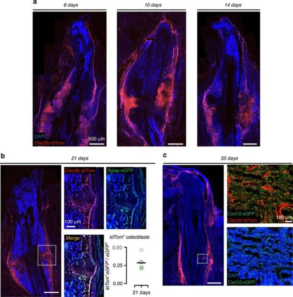
The team tagged these cells with a fluorescent label linked to Clec3b (a gene expressed by the inactive macrophage) using a mouse model. Under physiological conditions, Clec3b-positive cells remained localized to muscle and periosteum. After a breakthrough, they rushed to the site of injury.
By three weeks, about 28% of the new bone-forming osteoblasts within the healing callus came from this lineage. Others differentiated into bone marrow stromal cells that helped re-establish the support network within the bone.
Bones respond positively to external forces
But single-cell RNA sequencing confirmed the reprogramming: dormant progenitors switched to populations with molecular signatures of bone-forming and marrow-supportive cells. The key finding was that skeletal muscle served as a primary source of production.
Even when the periosteum was removed, muscle-derived FAPs still reached the fracture site. Bone grafts with surrounding muscle produced far more regenerative cells than grafts without muscle.
The same cells, however, also contributed to heterotopic ossification, abnormal bone growth in muscle and soft tissue after injury. Blocking key bone-forming pathways or depleting these cells reduced both fracture healing and unwanted bone formation.
“Our findings show that extra-skeletal cells, particularly FAPs, are recruited to help repair bone fractures and could represent a promising therapeutic target to enhance fracture healing,” said Dr. Ayturk.
This approach is tantalizing because these cells are responsible for both healing fractures and also contributing to the abnormal bone growth so obvious in Paget’s disease. Using them could expedite recovery, and controlled mobilization might curtail complications.
It reframes muscle not merely as a scaffolding structure for bones, but rather an underappreciated source of regenerative capacity, cells on standby ready to spring into action when injury calls.
Journal Reference:
- Aydin, E., Moore, J.A., Bubnovich, M. et al. Clec3b+ extraskeletal cells regulate fracture healing and heterotopic ossification. Bone Res 14, 70 (2026). DOI: 10.1038/s41413-026-00532-6


