
In recent times, scientists have made a groundbreaking discovery concerning inner-ear health. This research explores the puzzling issue of inner-ear bone loss, causing concern among medical professionals and researchers. Understanding this phenomenon could be key to developing effective treatments and preventative measures for related hearing and balance problems.
Chronic inflammation in the middle ear can lead to various issues and complications, affecting a person’s hearing and balance. One problem that can arise is the development of a cholesteatoma, an abnormal cell collection in the ear that can erode bones if not treated. This can cause hearing loss, dizziness, facial paralysis, and even brain infections.
Recently, scientists from Osaka University discovered the cause of cholesteatomas through a study published in Nature Communications, offering hope for developing new treatments for patients with this condition.
Cholesteatomas are abnormal growths in the ear made up of skin, collagen fibers, and other tissues. These growths can cause bone erosion, leading to hearing and balance problems. Scientists have been studying how cholesteatomas form.
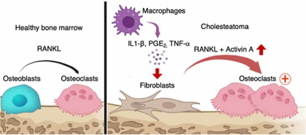
This new study from Osaka University used human tissues to identify cells called osteoclastogenic fibroblasts that trigger bone erosion. These cells produce a molecule called activin A, which causes bone breakdown and absorption by the body. Understanding this process may help develop better treatments for cholesteatomas.
Lead author Kotaro Shimizu said, “A cholesteatoma can still return or happen again even after its surgical removal, so it is important to know what is causing it.”
The researchers found a link between a molecule called activin A and bone erosion in cholesteatoma. According to senior author Masaru Ishii, targeting activin A could be a potential treatment for managing cholesteatomas. Currently, surgical removal is the only effective treatment for cholesteatomas. But this study’s discovery opens up the possibility of developing new medical treatments as the first-line management for cholesteatomas, providing hope for better therapies in the future.
In this study, the researchers investigated cholesteatoma, characterized by chronic middle ear inflammation and bone destruction. They analyzed human samples using single-cell RNA sequencing (scRNA-seq). They found a subset of fibroblasts expressing a molecule called INHBA/activin A, induced by proinflammatory cytokines.
Additionally, they established a mouse model of cholesteatoma. They observed that these pathogenic fibroblasts were responsible for ectopic osteoclastogenesis, the formation of bone-destroying cells. The researchers proposed a molecular mechanism for bone destruction in cholesteatoma, where proinflammatory cytokines induce activin A-expressing fibroblasts, which, in conjunction with RANKL, promote the formation of bone-destroying cells. The results suggest that targeting activin A and inflammatory cytokines may be potential therapeutic strategies for cholesteatoma treatment.
Journal Reference:
- Shimizu, K., Kikuta, J., Ohta, Y. et al. Single-cell transcriptomics of human cholesteatoma identifies an activin A-producing osteoclastogenic fibroblast subset inducing bone destruction. Nature Communications. DOI: 10.1038/s41467-023-40094-3.