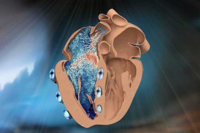
The right ventricle is one of the heart’s four chambers. It pumps deoxygenated blood to the lungs, so it doesn’t have to pump as hard. It’s a thinner muscle with more complex architecture and motion.
The intricate anatomy of the right ventricle has posed challenges for clinicians in accurately observing and assessing its function in patients with heart disease. Traditional tools frequently need to catch up in capturing the intricate mechanics and dynamics of the right ventricle, which can result in potential misdiagnoses and inadequate treatment strategies.
MIT engineers have developed a robotic replica of the heart’s right ventricle to improve understanding of the lesser-known chamber and speed the development of cardiac devices to treat its dysfunction. This robotic right ventricle, or RRV, mimics live hearts’ beating and blood-pumping action.
The robot ventricle integrates actual heart tissue with synthetic, balloon-like artificial muscles. This combination allows scientists to control the ventricle’s contractions while observing the functioning of its natural valves and other intricate structures.
The artificial ventricle can be adjusted to replicate both healthy and diseased states. The researchers manipulated the model to simulate conditions associated with right ventricular dysfunction, such as pulmonary hypertension and myocardial infarction. Additionally, they used the model to assess cardiac devices. For example, the team implanted a mechanical valve to repair a malfunctioning natural valve and observed how the ventricle’s pumping changed in response.
According to engineers, this RRV can be used as a realistic platform to study right ventricle disorders and test devices and therapies to treat those disorders. Plus, it can be used to study the effects of mechanical ventilation on the right ventricle and to develop strategies to prevent right heart failure in these vulnerable patients.
In a recent study, the team surgically removed a pig’s right ventricle, taking care to preserve its internal structures. They then applied a silicone wrapping around it, functioning as a soft, synthetic myocardium or muscular lining. Within this lining, several long, balloon-like tubes were embedded, encircling the actual heart tissue at positions optimized through computational modeling to replicate the ventricle’s contractions. Each tube was connected to a control system, allowing the researchers to inflate and deflate them at rates mimicking the heart’s natural rhythm and motion.
The team filled the model with a liquid viscosity similar to blood to evaluate its pumping capability. This transparent liquid allowed engineers to use an internal camera to observe how internal valves and structures responded as the ventricle pumped liquid through.
The study revealed that the artificial ventricle exhibited pumping power and internal structure functionality akin to what the scientists had observed in live, healthy animals. This demonstrates that the model can realistically simulate the action and anatomy of the right ventricle. Furthermore, the scientists were able to adjust the frequency and power of the pumping tubes to replicate various cardiac conditions, including irregular heartbeats, muscle weakening, and hypertension.
Roche said, “We’re reanimating the heart, in some sense, and in a way that we can study and potentially treat its dysfunction.”
To demonstrate the artificial ventricle’s capability in testing cardiac devices, the scientists surgically implanted ring-like medical devices of various sizes to repair the tricuspid valve of the chamber. The tricuspid valve is a leafy, one-way valve that allows blood into the right ventricle. When this valve is leaky or compromised, it can lead to right heart failure, atrial fibrillation, and symptoms such as reduced exercise capacity, leg and abdominal swelling, and liver enlargement.
The scientists surgically manipulated the robot-ventricle’s valve to simulate this condition and replaced it by implanting a mechanical valve or repairing it using ring-like devices of different sizes. They observed the impact of each device on the ventricle’s fluid flow as it continued to pump.
Manisha Singh, a postdoc at MIT’s Institute for Medical Engineering and Science (IMES), said, “With its ability to accurately replicate tricuspid valve dysfunction, the RRV serves as an ideal training ground for surgeons and interventional cardiologists. They can practice new surgical techniques for repairing or replacing the tricuspid valve on our model before performing them on actual patients.”
Journal Reference:
- Singh, M., Bonnemain, J., Ozturk, C. et al. Robotic right ventricle is a biohybrid platform that simulates right ventricular function in (patho)physiological conditions and intervention. Nat Cardiovasc Res 2, 1310–1326 (2023). DOI: 10.1038/s44161-023-00387-8
