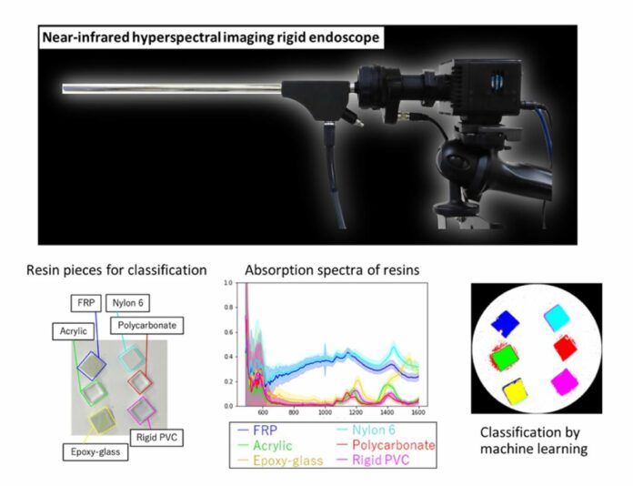
The food and industrial sectors have shown interest in near-infrared hyperspectral imaging (NIR-HSI), which obtains spectral images in the NIR region (850–2500 nm). The components of objects that are difficult to differentiate with visible light, such as the fat content of meat, the makeup of resins with similar colors, and impurities from foreign matter, can be nondestructively analyzed using this imaging technology.
Over-thousand-nanometer (OTN) spectroscopy is a noteworthy feature of NIR-HSI that can be used for 2D map construction, organic chemical identification, and concentration estimate. Moreover, NIR-HSI helps visualize tumors concealed in healthy tissues since it can gather data deep within the body.
Various types of HSI devices exist based on different imaging targets and situations. But for OTN wavelengths, ordinary visible cameras lose sensitivity, and only a few commercially available lenses can correct chromatic aberration.
In a new study, scientists from Tokyo University of Science have developed the world’s first rigid endoscope system capable of HSI from visible to OTN wavelengths. This new system can perform hyperspectral imaging (HSI) between visible and 1600 nm wavelengths using a supercontinuum light source and an acousto-optic tunable filter to emit specific wavelengths.
The system comprises a Supercontinuum (SC) light source and an acoustic-opto tunable filter (AOTF) that can emit specific wavelengths. This combination allows simple light transmission to the light guide and millisecond-fast electrical switching between various wavelengths.
The system’s optical performance was verified, and the classification ability was investigated. Consequently, it was demonstrated that HSI (490–1600 nm) could be performed.
Advantages:
- The low light power of extracted wavelengths.
- Non-destructive imaging and downsizing capability.
- A more continuous NIR spectrum can be obtained.
Scientists demonstrated the system’s capability by getting the spectra of six types of resins. They employed a neural network to classify the spectra pixel-by-pixel in multiple wavelengths.
The findings showed that the neural network could categorize seven distinct targets, comprising the six resins and a white reference, with an accuracy of 99.6%, repeatability of 93.7%, and specificity of 99.1% when the OTN wavelength range was taken from the HSI data for training. This indicates that the system may extract each resin pixel’s molecular vibration information successfully.
Professor Hiroshi Takemura from Tokyo University of Science (TUS) said, “This breakthrough, which combines expertise from different fields through a collaborative, cross-disciplinary approach, enables the identification of invaded cancer areas and the visualization of deep tissues such as blood vessels, nerves, and ureters during medical procedures, leading to improved surgical navigation. Additionally, it enables measurement using light previously unseen in industrial applications, potentially creating new areas of non-use and non-destructive testing. By visualizing the invisible, we aim to accelerate the development of medicine and improve the quality of life of physicians and patients.”
Journal Reference:
- Toshihiro Takamatsu, Ryodai Fukushima, Kounosuke Sato, Masakazu Umezawa, Hideo Yokota, Kohei Soga, Abian Hernandez-Guedes, Gustavo M. Callico, and Hiroshi Takemura. Development of a visible to 1600 nm hyperspectral imaging rigid-scope system using supercontinuum light and an acousto-optic tunable filter. Optics Express. DOI: 10.1364/OE.515747
