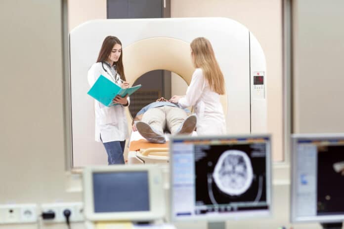
A team of UC Davis researchers has made a significant breakthrough by using dynamic total-body positron emission tomography (PET) to capture the human body’s immune response to COVID-19 in recovering patients. Their findings, published in Science Advances, may improve our understanding of how the immune system fights viral infections and provides long-term protection against reinfection.
The researchers utilized the Explorer total-body PET scanner, a state-of-the-art imaging technology developed in partnership with United Imaging Healthcare. Dynamic PET involves administering tiny amounts of radiotracers and continuously monitoring their distribution over time in the body. This dynamic imaging process resembles a movie and enables the extraction of valuable biological information using mathematical models.
Total-body PET scanners allow dynamic imaging and modeling of all body organs simultaneously. These scanners offer much greater sensitivity compared to standard PET systems. This results in more explicit images while reducing the radiotracer needed for the scan.
First author Negar Omidvari, an assistant project scientist in the UC Davis Department of Biomedical Engineering, said, “Dynamic total-body PET is currently the only available technology with an acceptable radiation dose that allows noninvasive quantitative measurements of immune cell distribution and trafficking (movement) inside all tissues in living humans.”
This study marks the first use of dynamic PET and kinetic modeling to track CD8+ T cells in humans. These cells, equipped with CD8 proteins on their surface, play a crucial role in responding to viral infections. They become active and hunt down infected cells, providing short-term protection. Some transform into memory T cells, which offer long-lasting defense against reinfection.
While these cells travel in the bloodstream, they mainly work in non-blood tissues, like the bone marrow, spleen, tonsils, and lymph nodes.
Omidvari said, “There has been a growing interest in studying the critical role of CD8+ T cells in immune response and memory. However, evaluating immunological changes in non-blood tissue is challenging due to the invasive nature of biopsies. In some cases, it is not even practical in certain anatomical regions of living participants, such as the brain, spinal cord, cardiopulmonary tissue, and vascular tissue. So, the challenge was to find noninvasive quantitative methods for measuring CD8+ T cell distribution and trafficking in the body that is safe to also use in healthy people.”
The study involved three healthy individuals and five patients recovering from mild to moderate COVID-19 (not hospitalized). Researchers gave them a small dose of a radioactive liquid containing an immunoPET radiotracer (89Zr-Df-Crefmirlimab) that targets human CD8.
Each participant had three scans: one lasting 90 minutes, another at six hours, and a final one at 48 hours after the injection. Recovering patients repeated the same scans four months later.
The researchers assessed the radiotracer’s activity in both blood and non-blood tissues in the PET images. They used kinetic modeling to separate the influence of blood flow on the tissues. This allowed them to gauge radiotracer absorption in the tissues independently of the scan’s timing and individual blood variations.
Using total-body PET, the researchers achieved noninvasive T-cell distribution measurements with exceptional image quality across the entire body and all types of tissues. Their study revealed a strong presence of CD8+ T cells in the lymphoid organs of all participants, with the highest concentration in the spleen, followed by the bone marrow, liver, tonsils, and lymph nodes.
The most significant discovery was the higher levels of CD8+ T cells in the bone marrow of recovering COVID-19 patients compared to healthy individuals. In follow-up scans taken six months after infection, these levels in recovering patients remained slightly higher than the baseline measurements taken about two months post-infection.
This suggests that the bone marrow serves as a significant reservoir and a preferred site for the proliferation of memory CD8+ T cells following a viral infection. The migration of memory T cells to specific tissues, such as the bone marrow, plays a critical role in developing immune memory after viral infections.
The combination of a precise immunoPET tracer and the exceptional detection sensitivity of total-body PET creates an innovative platform for scientists. It allows them to explore the immune response and memory in all organs over extended periods without invasive methods.
Simon R. Cherry, a UC Davis physicist, emphasizes the study’s significance. It showcases the potential of total-body PET for assessing T-cell distribution throughout the entire human body. The imaging quality supports detailed modeling, and the low radiation dose makes it suitable for widespread use in studying the human immune response. This research characterizes the dynamics of the immune tracer in healthy subjects and those with infectious diseases, such as COVID-19, marking a significant milestone.
The researchers highlighted numerous potential applications for their method. It can be applied to investigate the immune response in viral infections, understand immune memory post-infection, and assess treatment responses in cancer patients. This versatile method may also find utility in studying infectious diseases, autoimmune disorders, and organ transplants. Additionally, it can be used in prognosis, therapeutic development, and vaccine research.
Journal reference:
- Negar Omidvari, Terry Jones, et al., First-in-human immunoPET imaging of COVID-19 convalescent patients using dynamic total-body PET and a CD8-targeted minibody. Science Advances. DOI: 10.1126/sciadv.adh7968.
