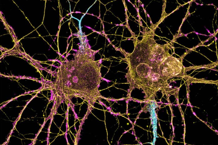
Huntington’s disease is a rare, inherited disease that causes the progressive breakdown (degeneration) of nerve cells in the brain. In patients with Huntington’s disease, Striatal projection neurons (SPNs) progressively degenerates, contributing to patients’ loss of motor control, which is one of the significant hallmarks of the disease.
It is well established that the indirect-pathway SPNs are susceptible to neurodegeneration and transcriptomic disturbances. Still, less is known about how the striosome-matrix axis is compromised in HD to the canonical axis.
Neuroscientists at MIT have now established that Huntington’s disease has diverse effects on two different cell groups in the striatum. They postulate that motor impairments result from the neurodegeneration of one of these populations. Still, the mood abnormalities frequently observed in the early stages of the disease may be caused by damage to the other population, which is found in structures called striosomes.
Scientists used single-cell RNA sequencing to analyze the genes expressed in mouse models of Huntington’s disease and postmortem brain samples from Huntington’s patients. They found that the cells of the striosomes and another structure, the matrix, begin to lose their distinguishing features as the disease progresses.
Scientists noted, “This analysis could also shed light on other brain disorders that affect the striatum, such as Parkinson’s disease and an autism spectrum disorder.”
Neurons in the striatum can be categorized as D1 or D2 neurons. D1 neurons play a role in the “go” pathway, which starts an action, while D2 neurons play a role in the “no-go” pathway, which stops an action. The striosomes and the matrix are both home to D1 and D2 neurons.
Striosomal neurons are more severely affected by Huntington’s disease than matrix neurons, according to an examination of RNA expression in each type of cell. Furthermore, D2 neurons are more susceptible than D1 neurons inside the striosomes.
Scientists also demonstrate that these four major cell types begin to lose their identifying molecular identities and become more difficult to distinguish from one another in Huntington’s disease. In other words, the distinction between striosomes and matrix becomes blurry, noted scientists.
Ann Graybiel, an MIT Institute Professor and a member of MIT’s McGovern Institute for Brain Research, said, “The findings suggest that damage to the striosomes, which are known to be involved in regulating mood, may be responsible for the mood disorders that strike Huntington’s patients in the early stages of the disease. Later on, degeneration of the matrix neurons likely contributes to the decline of motor function.”
Scientists are looking forward to exploring how degeneration or abnormal gene expression in the striosomes may contribute to other brain disorders.
Graybiel said, “There are many, many disorders that probably involve the striatum, and now, partly through transcriptomics, we’re working to understand how all of this could fit together.”
Journal Reference:
- Matsushima, A., Pineda, S.S., Crittenden, J.R. et al. Transcriptional vulnerabilities of striatal neurons in human and rodent models of Huntington’s disease. Nat Commun 14, 282 (2023). DOI: 10.1038/s41467-022-35752-x
