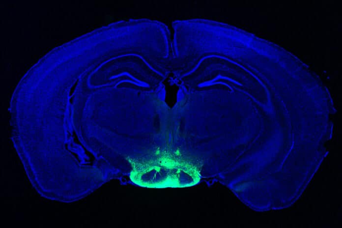
The neural circuits governing the induction and progression of neurodegeneration and memory impairment in Alzheimer’s disease (AD) are incompletely understood. n a new study, MIT researchers have identified a subset of neurons within the mammillary body that is most susceptible to neurodegeneration and hyperactivity.
According to the study, this region is a promising target for potential new medications to treat Alzheimer’s disease because it may be involved in some of its first symptoms.
In Alzheimer’s disease, only the lateral mammillary body neurons experience neurodegeneration and hyperactivity. The medial mammillary body neurons do not. Using a medication already used to treat epilepsy, the researchers demonstrated in a mice trial that they could repair memory impairments brought on by hyperactivity and neurodegeneration in mammillary body neurons.
In 2019, scientists used a mouse model of Alzheimer’s disease. They identified that the mammillary bodies had the highest density of amyloid beta. These bodies are known to be involved in memory, but their exact role in normal memory and Alzheimer’s disease is unknown.
The researchers employed single-cell RNA sequencing, which can show the active genes within different types of cells in a tissue sample, to learn more about the function of the mammillary body. With this method, they could distinguish between two important groups of neurons: one in the lateral and one in the medial mammillary bodies. The researchers discovered that the lateral neurons exhibited higher spiking rates than the medial mammillary body neurons and that synaptic activity-related genes were strongly expressed in these neurons.
The researchers questioned whether the lateral neurons would be more prone to Alzheimer’s disease in light of these changes. They looked at a mouse model with five genetic variants associated with human early-onset Alzheimer’s disease to answer that question. The scientists discovered that compared to healthy mice, these mice displayed significantly higher lateral mamillary body neuron hyperactivity. However, no such alterations existed in the medial mammillary body neurons of healthy mice and those in the Alzheimer’s model.
The scientists discovered that before amyloid plaques started to form, this hyperactivity appeared exceptionally early, at roughly two months of age (the equivalent of a young adult human). As the mice grew older, the lateral neurons became even more excitable, and they were also more vulnerable to neurodegeneration than the medial neurons.
Murdock says, “We think the hyperactivity is related to dysfunction in memory circuits and is also related to a cellular progression that might lead to neuronal death.”
In the Alzheimer’s mouse model, new memory formation was impaired, but when the mice were given a medication that lessens neuronal excitation, their performance on memory tests was noticeably improved. Levetiracetam, a medication used to treat epileptic seizures, is also being tested in clinical settings for the treatment of Alzheimer’s patients’ epileptiform activity, a condition marked by hyperexcitability in the cortex that raises the risk of seizures.
The Religious Orders Study/Memory and Ageing Project (ROSMAP), a longitudinal study that has tracked memory, motor, and other age-related disorders in senior citizens since 1994, was also used to analyze human brain tissue. The researchers discovered two clusters of neurons that correlate to the lateral and medial mammillary body neurons found in mice using single-cell RNA-sequencing of mammillary body tissue from persons with and without Alzheimer’s disease.
The researchers discovered signs of hyperactivity in the lateral mammillary bodies from Alzheimer’s tissue samples, including upregulation of genes that encode potassium and sodium channels. These signs were similar to those found in the mice experiments. In those samples, they also discovered that the lateral neuron cluster had more neurodegeneration than the medial cluster.
Scientists noted, “Other studies of Alzheimer’s patients have found a loss of volume of the mammillary body early in the disease, along with deposition of plaques and altered synaptic structure. All of these findings suggest that the mammillary body could make a good target for potential drugs that could slow down the progression of Alzheimer’s disease.”
Journal Reference:
- Wen-Chin Huang, Zhuyu Peng, Mitchell H. Murdock et al. Lateral mammillary body neurons in the mouse brain are disproportionately vulnerable to Alzheimer’s disease. Science Translational Medicine. DOI: 10.1126/scitranslmed.abq1019
