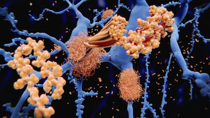
Amyloid fibrils are self-assembled fibrous protein aggregates that are associated with several presently incurable diseases such as Alzheimer’s and Parkinson’s disease.
Amyloid fibrils are shaped by the notorious amyloid-beta peptide and Tau protein, which are two of the most sought-after targets for the development of therapies to treat Alzheimer’s and similar diseases.
The fibril structures spread throughout the brain by moving from one cell to another. This is thought to lead to the degeneration of neurons, causing brain damage and cognitive impairments like memory loss.
Given the significance of amyloid fibrils, there have been numerous endeavors to imagine them in however much detail as could reasonably be expected to gain detailed insights about their structure. Unraveling their structural details might prompt detection of weak spots that could be targeted for treatment and pave the way for developing more reliable diagnostic tools. Despite much work, however, imaging and capturing the diversity of fibrils in biological samples has proven to be very difficult due to their complex nature and heterogeneity.
In a new study by the groups of Francesco Stellacci and Hilal Lashuel at EPFL, scientists have discovered a solution. They have shown that gold amphiphilic anionic nanoparticles with a diameter around 3 nm could effectively label the edge of amyloid fibrils in a hydrated state. This makes the visualization of the diverse amyloid fibrils easier.
Scientists imaged the nanoparticles-decorated fibrils using a specialized form of TEM called “cryogenic transmission electron microscopy” (cryo-EM). They detected the main difference that in cryo-EM, the sample – here the fibrils – is first rapidly frozen to a very low temperature and can be visualized in its “natural” state without having to be fixed or stained beforehand.
Between the profoundly efficient binding of gold nanoparticles and the capabilities of cryo-EM, the acquired images of fibrils and unmask their diversity with exceptional clarity. This included fibrils developed in the lab just as from actual postmortem tissues of patients.
Stellacci said, “Our findings reveal a striking morphological difference between the fibrils produced in cell-free systems and those isolated from patients. This supports the current view that the physiologic environment plays a major role in determining different types of amyloid fibrils.”
Lashuel said, “These advances pave the way for elucidating the structural basis of amyloid strains and toxicity. The nanoparticles are powerful and desperately needed tools for rapid imaging and profiling of amyloid morphological polymorphism in different types of samples under cryo-conditions, especially complex samples isolated from human-derived pathological aggregates.”
The study is published in the journal PNAS.
