Scintillators are materials that can alter high-energy radiation to a near-visible or visible light. This scintillation process is used in many detector applications ranging from medical imaging to high-energy particle physics.
There is great interest in developing better scintillators with greater photon yields and improved spatial and energy resolution. Most studies that focus on improving scintillators involve synthesizing new materials with better intrinsic scintillating properties.
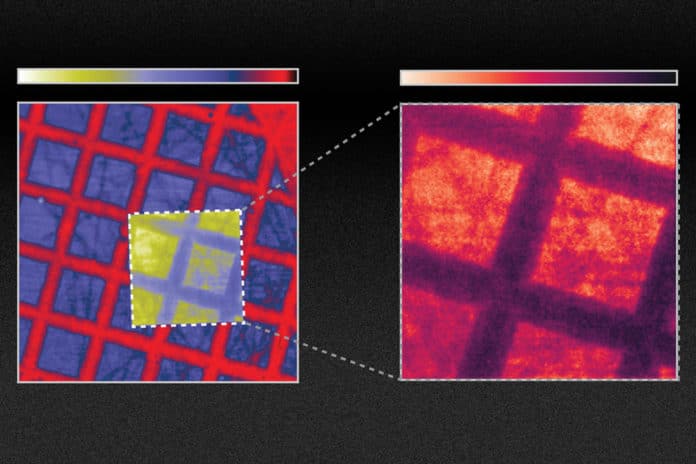
MIT scientists have now shown that one could improve the efficiency of scintillators by 10x to 100x. It can be done by changing the material’s surface to create specific nanoscale configurations, such as arrays of wave-like ridges.
Instead of developing new materials that produce brighter or faster light emissions, this new approach applies advances in nanotechnology to existing materials.
Scientists created a pattern of holes in scintillator materials at a length scale comparable to the wavelengths of the light being emitted. The holes were separated by roughly one optical wavelength or about 500 nanometers. Doing so prompted a drastic change in the material’s optical properties.
Roques-Carmes said, “To make what they coined nanophotonic scintillators, you can directly make patterns inside the scintillators, or you can glue on another material that would have holes on the nanoscale. The specifics depend on the exact structure and material.”
Thanks to a general theory and the framework that scientists developed, scientists were able to calculate the scintillation levels produced by any arbitrary configuration of nanophotonic structures.
Roques-Carmes said, “The scintillation process itself involves a series of steps, making it complicated to unravel. The framework the team developed involves integrating three different types of physics. Using this system, we found a good match between their predictions and the results of their subsequent experiments.”
“The experiments showed a tenfold improvement in emission from the treated scintillator. So, this might translate into applications for medical imaging, which are optical photon-starved, meaning the conversion of X-rays to optical light limits the image quality. [In medical imaging,] you do not want to irradiate your patients with too much of the X-rays, especially for routine screening, and especially for young patients as well.”
“We believe that this will open a new field of research in nanophotonics. You can use a lot of the current work and research that has been done in the field of nanophotonics to improve significantly on existing materials that scintillate.”
Rajiv Gupta, chief of neuroradiology at Massachusetts General Hospital and an associate professor at Harvard Medical School, said, “The research presented in this paper is hugely significant. Nearly all detectors used in the $100 billion [medical X-ray] industry are indirect detectors, which is the type of detector the new findings apply to.”
“Everything that I use in my clinical practice today is based on this principle. This paper improves the efficiency of this process by ten times. If this claim is even partially true, say the improvement is two times instead of 10 times, it would be transformative for the field!”
Journal Reference:
- Charles Roques-Carmes et al. A framework for scintillation in nanophotonics. DOI: 10.1126/science.abm9293
