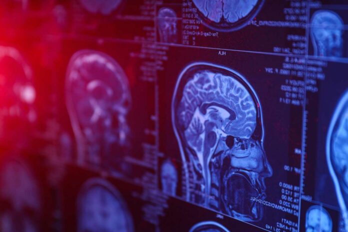
MIT and Harvard researchers use advanced microscopy to see hidden structures in the human brain, revealing surprising details about low-grade tumors. The technique aims for better tumor diagnosis, accurate prognosis, and improved treatment decisions.
Pablo Valdes, a former MIT postdoc who is now an assistant professor of neuroscience at the University of Texas Medical Branch and the lead author of the study, said, “We’re starting to see how important the interactions of neurons and synapses with the surrounding brain are to the growth and progression of tumors. A lot of those things we really couldn’t see with conventional tools, but now we have a tool to look at those tissues at the nanoscale and try to understand these interactions.”
MIT’s Edward Boyden and Harvard’s E. Antonio Chiocca developed an imaging method using expansion microscopy, making molecules visible in high resolution without expensive microscopes. The study is published in Science Translational Medicine.
The method involves
- embedding tissue in a swelling polymer,
- breaking protein bonds with water and
- expanding it.
This allows high-resolution images, previously only possible with expensive microscopes. In 2017, Boyden’s lab expanded preserved human tissue, but the chemicals destroyed labeled proteins. The new approach uses fluorescent antibodies before expansion, enabling the visualization of proteins’ location and identity post-expansion.
In this study, researchers developed a new method to soften tissue, preserving proteins. After expansion, fluorescent antibodies label proteins. Multiple imaging rounds are possible, allowing observation of structures in crowded spaces. MIT Assistant Professor Deblina Sarkar demonstrated this ‘decrowding’ technique in 2022 using mouse tissue.
The study introduces a ‘decrowding’ technique for human brain tissue in clinical samples. Over 16 molecules were labeled, including markers for structures and cell types and those linked to tumor aggressiveness and neurodegeneration. The approach analyzed healthy and glioma-afflicted brain tissues, providing valuable insights for pathology and treatment decisions.
Valdes said, “We wanted to look at brain tumors to understand them better at the nanoscale level and, by doing that, to develop better treatments and diagnoses. At this point, it was more developing a tool to understand them better because currently, in neuro-oncology, people haven’t done much in super-resolution imaging.”
Researchers labeled vimentin, a protein found in highly aggressive glioblastomas, to spot aggressive cells in gliomas. Surprisingly, they found more vimentin-expressing cells in low-grade gliomas than traditional methods showed.
This insight into tumor biology suggests that some tumors may be more aggressive than standard staining techniques indicate. Typically, during glioma surgery, preserved tumor samples are analyzed using immunohistochemistry staining, revealing markers of aggressiveness, including those examined in this study.
The discovery aids in targeting molecules for better treatments of incurable brain cancers. The collaboration between clinicians and scientists aims to enhance patients’ lives. The expansion microscopy technique could provide more insights into tumors, determining aggressiveness and guiding treatment choices.
A more extensive study is planned to establish diagnostic guidelines based on revealed tumor traits, hoping to make it a practical tool for neuro-oncology and neuropathology in examining neurological diseases at the nanoscale.
Boyden’s lab plans to use the nanoimaging technique to study various aspects of brain function, both in healthy and diseased tissue. Nanoimaging is crucial for understanding nanoscale interactions in biology, such as genes, gene products, and biomolecules.
The research, funded by various organizations, including the Howard Hughes Medical Institute and the Bill and Melinda Gates Foundation, explores synaptic changes, immune interactions, and alterations during cancer and aging.
Journal reference:
- PABLO A. VALDES , CHIH-CHIEH (JAY) YU et al., Improved immunostaining of nanostructures and cells in human brain specimens through expansion-mediated protein decrowding. Science Translational Medicine. DOI: 10.1126/scitranslmed.abo0049.
