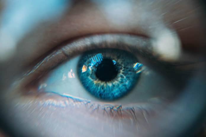
In vivo observation of the human retina at the cellular level is crucial to detect the first signs of retinal diseases and adequately treat them. Despite the phenomenal advances in adaptive optics systems, clinical imaging of many retinal cells is still elusive due to the low signal-to-noise ratio induced by transpupillary illumination.
Scientists at the Laboratory of Applied Photonics Devices (LAPD), have developed a device that can zoom in on previously invisible cells at the back of the eye.
The device uses a sophisticated imaging system to view the macula cell layers – the first ones affected by AMD – in real-time. It looks at the retina through the sclera, which is the white of our eyes.
Mathieu Künzi, a researcher at the LAPD and co-author of the study, said, “This means we get to see the back of the eye from a different, diagonal angle. That prevents some of the interference that can come from reflected light and gives us a better view of the cell layers.”
Mathieu Künzi, along with his colleague Timothé Laforest, another LAPD researcher, has created a startup – EarlySight – to develop and promote this technology in the medical world.
When tested on a dozen healthy people, scientists found that the device is reliable and ten times more accurate in observing the back of the eye than conventional methods. It can show the different stages that those cells go through, particularly during the aging process.
The device is developed in partnership with the Faculty of Biology and Medicine, University of Lausanne, Hôpital Jules-Gonin, Lausanne, Centre de Recherche des Cordeliers in Paris, Ophthalmology Department of Hôpital Cochin in Paris, EarlySight SA.
Journal Reference:
- Transscleral optical phase imaging of the human retina. DOI: 10.1038/s41566-020-0608-y
