
Researchers at the Center for Quantum Nanoscience (QNS) within the Institute for Basic Science (IBS) at Ewha Womans University in collaboration with colleagues from the US have performed the world’s smallest magnetic resonance imaging to capture the magnetic fields of single atoms. This is an incredible achievement that can improve quantum research, as well as our perception of the universe on a subatomic scale.
Magnetic Resonance Imaging (MRI) machines are great for creating detailed images of the inside of a person’s body. Using strong magnetic fields and radio waves, MRI’s detect the density of spins – the fundamental magnets in electrons and protons – in the human body, all without producing side-effects.
The machines temporarily change how the billions of protons in the person’s body spin. Then they measure and image energy released by these protons once they return to their normal state. But for medical MRI scanners, billions of protons are required for the sensors to detect it.
The team of researchers shows that this process is now also possible for an individual atom on a surface. To bring the process down to much finer scales, the team used a Scanning Tunneling Microscope – which consists of an atomically sharp metal tip – that can image surfaces at the atomic scale by running an extremely fine needle over them.
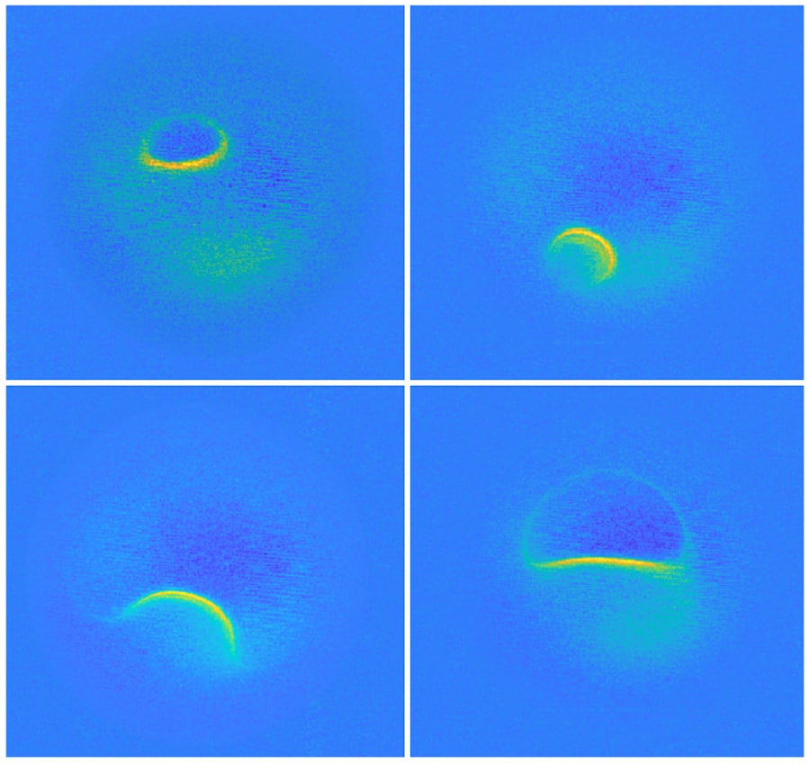
For their study, the team swept the microscope’s tip over iron and titanium atoms they’d placed on a magnesium oxide surface. This puts the atoms to a magnetic field that disrupted their electrons. Researchers then hit the atoms with a radio wave pulse, and the system imaged the energy the electrons subsequently released.
“It turns out that the magnetic interaction we measured depends on the properties of both spins, the one on the tip and the one on the sample,” says lead author, Dr. Philip Willke at the Center for Quantum Nanoscience (QNS). “For example, the signal that we see for iron atoms is vastly different from that for titanium atoms. This allows us to distinguish different kinds of atoms by their magnetic field signature and makes our technique very powerful.”
The team is planning to use their single-atom MRI to map the spin distribution in more complex structures such as molecules and magnetic materials. “Many magnetic phenomena take place on the nanoscale, including the recent generation of magnetic storage devices,” says Dr. Yujeong Bae also of QNS, a co-author in this study. “We now plan to study a variety of systems using our microscopic MRI.”
They also believe that this new nanoscale imaging technique could lead to the development of new materials and drugs, as well as the creation of better quantum computing systems.
“The ability to map spins and their magnetic fields with previously unimaginable precision allows us to gain deeper knowledge about the structure of matter and opens new fields of basic research,” says Prof. Andreas Heinrich, Director of QNS.
The new findings are published today in the journal Nature Physics.