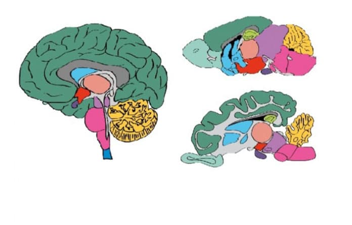
The brain is Of all the objects in the universe, the human brain is the most complex: There are as many neurons in the brain as there are stars in the Milky Way galaxy. But we are at least beginning to grasp the crucial mysteries of neuroscience and starting to make headway in addressing them.
The scientists at the Karolinska Institutet have put a new effort by launching a comprehensive overview of all proteins expressed in the brain. This overview offers medical researchers an unprecedented resource to deepen their understanding of neurobiology and develop new, more effective therapies and diagnostics targeting psychiatric and neurological diseases.
The new Brain Atlas resource depends on the study of almost 1,900 brain tests covering 27 brain regions, combining data from the human brain with relating data from the brains of the pig and mouse.
Mathias Uhlén, Professor at the Department of Protein Science at KTH Royal Institute of Technology, said, “As expected, the blueprint for the brain is shared among mammals, but the new map also reveals interesting differences between human, pig and mouse brains.”
Associated to psychiatric disorders
The cerebellum is the area at the back and bottom of the brain, behind the brainstem. This part emerged in the study as the most distinctive region of the brain. It has several functions relating to movement and coordination, including Maintaining balance.
The proteins in the cerebellum, especially those with elevated expression levels, are associated with psychiatric disorders, supporting the role of the cerebellum in the processing of emotions.
Dr. Evelina Sjöstedt, a researcher at the Department of Neuroscience at Karolinska Institutet, said, “Another interesting finding is that the different cell types of the brain share specialized proteins with peripheral organs. For example, astrocytes, the cells that ‘filter’ the extracellular environment in the brain, share a lot of transporters and metabolic enzymes with cells in the liver that filter the blood.”
Essential differences between the species
Neurotransmitters are often referred to as the body’s chemical messengers. They are the molecules used by the nervous system to transmit messages between neurons, or neurons to muscles.
Communication between two neurons happens in the synaptic cleft. Here, electrical signals that have traveled along the axon are briefly converted into chemical ones through the release of neurotransmitters, causing a specific response in the receiving neuron.
A comparison of neurotransmitter systems could reveal clear differences between the species.
Dr. Jan Mulder, group leader of the Human Protein Atlas brain profiling group, said, “Several molecular components of neurotransmitter systems, especially receptors that respond to released neurotransmitters and neuropeptides, show a different pattern in humans and mice. This means that caution should be taken when selecting animals as models for human mental and neurological disorders.”
Human Protein Atlas (HPA) program
Started in 2003, the Human Protein Atlas (HPA) program aims to map all of the human proteins in cells, tissues, and organs (the proteome).
The database also contains microscopic images for selected genes/proteins to show the protein distribution in human brain samples and detailed, zoomable maps of protein distribution in the mouse brain.
This overview is the latest database released by the Human Protein Atlas (HPA) program, which is based at the Science for Life Laboratory (SciLifeLab) in Sweden, joint research center-aligned with KTH Royal Institute of Technology, Karolinska Institutet, Stockholm University and Uppsala University. The project is a collaboration with the BGI research center in Shenzhen and Qingdao in China and Aarhus University in Denmark.
The database also contains microscopic images for selected genes/proteins to show the protein distribution in human brain samples and detailed, zoomable maps of protein distribution in the mouse brain.
The main funding for the research was provided by the Knut and Alice Wallenberg Foundation.
The publication is published in the journal Science.
