Passing out brief losses of consciousness- whether brought by pain, fear, heat, hyperventilation, or other causes- leads to hospital emergency room visits. However, the mechanisms behind this remain unknown.
A new study by University of California San Diego scientists, The Scripps Research Institute, and other institutions identified the genetic pathway between the heart and brain tied to fainting.
Considering the heart as a sensory organ, as opposed to the conventional wisdom that the brain sends signals and the heart merely obeys, was one of their novel strategies. Scientists in this study use various techniques to comprehend these neurological connections between the brain and heart.
School of Biological Sciences Assistant Professor Vineet Augustine, the paper’s senior author, said, “What we are finding is that the heart also sends signals back to the brain, which can change brain function. Our study is the first comprehensive demonstration of a genetically defined cardiac reflex, which faithfully recapitulates characteristics of human syncope at physiological, behavioral, and neural network levels.”
Scientists studied neural mechanisms related to the Bezold-Jarisch reflex (BJR), a cardiac reflex first described in 1867. For long, BJR, characterized by lowered blood pressure, respiration, and heart rate, is hypothesized to be linked to fainting. However, since the brain circuits involved in the response were poorly understood, data to support the theory was lacking.

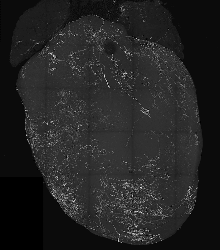
The nodose ganglia, a sensory cluster that is a component of the vagus nerves that transmit messages from the brain to visceral organs like the heart, are the subject of the study’s genetic focus. In particular, vagal sensory neurons, or VSNs, are believed to be connected to BJR and fainting. VSNs send signals to the brainstem. While searching for a new neurological pathway, scientists found a strong correlation between the well-known BJR responses and VSNs that express the neuropeptide Y receptor Y2 (NPY2R).
While investigating this route in mice, scientists discovered, to their astonishment, that mice that had been moving freely suddenly fainted when they proactively stimulated NPY2R VSNs using optogenetics, a technique for producing and controlling neurons. Thousands of neurons in the mice’s brains were recorded during these events, along with changes in the heart rate and facial traits like whisking and pupil diameter.
To evaluate the data and identify relevant aspects, they also used machine learning in several methods. They discovered that when NPY2R neurons were triggered, mice showed reduced heart rate, blood pressure, and breathing rate in addition to fast pupil dilation and the well-known “eye-roll” that occurs when a person faints. Additionally, as part of their partnership with Professor David Kleinfeld’s group in the UC San Diego Departments of Neurobiology and Physics, they evaluated decreased blood flow to the brain.
Scientists were surprised to see how their eyes rolled back around the same time as brain activity rapidly dropped. After a few seconds, brain activity and movement returned.
Scientists noted, “This was our eureka moment.”
Additional tests revealed that the BJR and fainting problems disappeared in mice whose NPY2R VSNs were eliminated. The current study confirmed the previous findings that a decrease in brain blood flow causes fainting, but it also suggested that brain activity itself may be a significant factor. Thus, the results indicate that the recently discovered genetically identified VSNs and associated neural pathways are activated not only in BJR but also more broadly in animal physiology, specific brain networks, and behavior.
Augustine said, “Such findings were difficult to tease out previously because neuroscientists study the brain and cardiologists study the heart, but many do so in isolation of the other. Neuroscientists traditionally think the body follows the brain, but now it is becoming obvious that the body sends signals to the brain, and then the brain changes function.”
The results of their study have led the researchers to want to keep observing the exact circumstances in which vagal sensory neurons fire.
Scientists noted, “We also hope to more closely examine cerebral blood flow and neural pathways in the brain during the moment of syncope to understand this common but mysterious condition better.”
Journal Reference:
- Lovelace, J.W., Ma, J., Yadav, S. et al. Vagal sensory neurons mediate the Bezold–Jarisch reflex and induce syncope. Nature (2023). DOI: 10.1038/s41586-023-06680-7
