
Scientists at the Imperial College London have recently come up with a new prototype device- Mini magic MRI scanner to detect injury such as partial ligament tears and to help diagnose knee injuries more quickly and precisely.
When tested on animal knees, the outcomes suggest that the technology could be used to show all the structures of the knee.
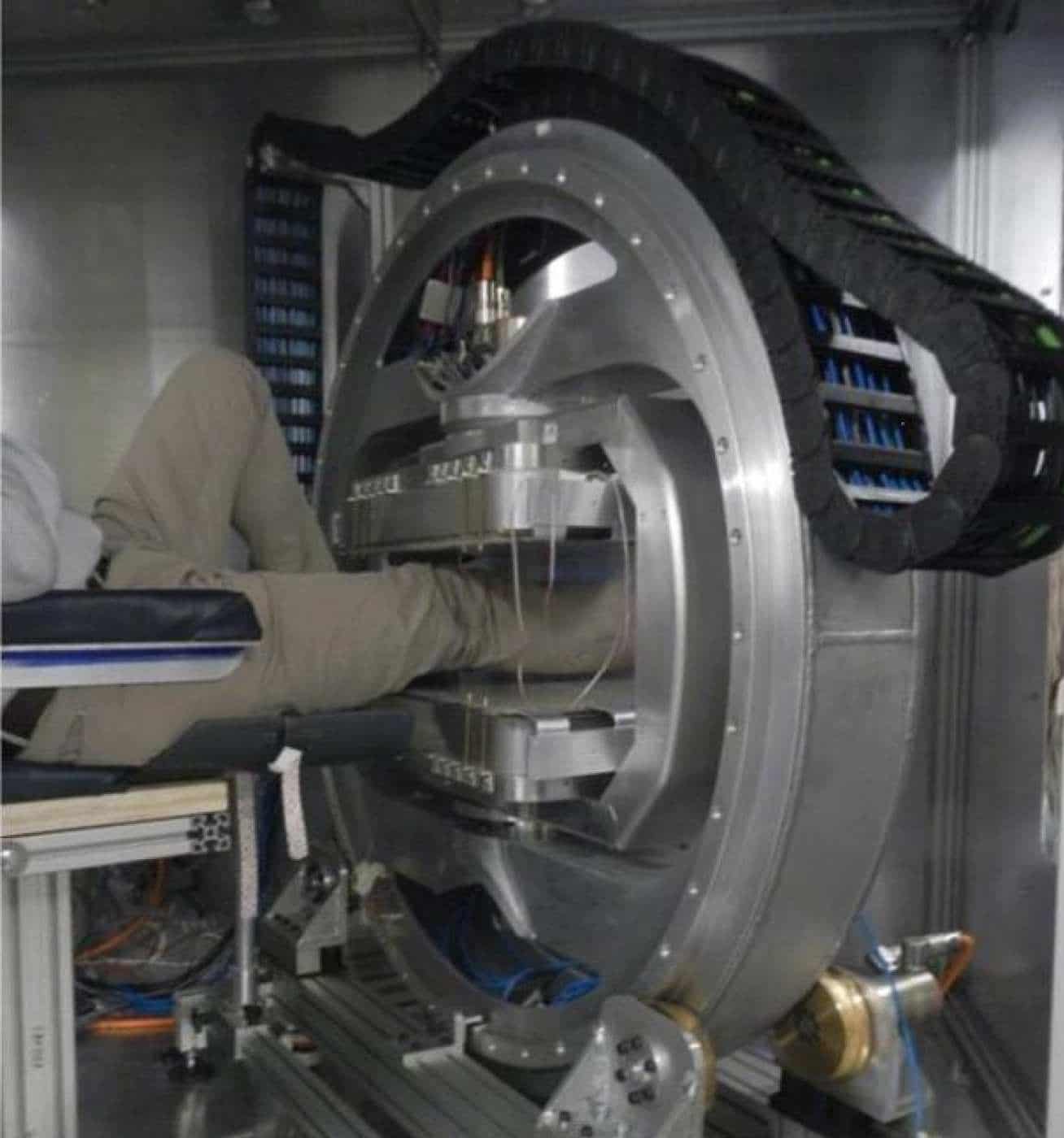
The prototype device comes in small size. Thus it can be used in local clinics and even GP surgeries, potentially reducing NHS waiting times for MRI scans.
Dr. Karyn Chappell, a researcher, and radiographer from Imperial’s MSK Lab said, “Currently, key components of the knee joints such as ligaments and tendons are difficult to see in detail in the MRI scans. Knee injuries affect millions of people – and MRI scans are crucial to diagnosing the problem, leading to quick and effective treatment. However, we currently face two problems: connective tissue in the knee is unclear on MRI scans, and people are waiting a long time for a scan.”
“This can cause particular problems for women, as they are at greater risk of anterior cruciate ligament injuries. The reasons for this are unclear, but it could be linked to hormones such as estrogen making ligaments more elastic, leading to more joint injuries.”
Following knee damage, a doctor may refer a patient for an MRI scan to help establish up which part portion of the joint is injured. MRI scans use a combination of radio waves and strong magnets to ‘flip’ water molecules in the body. The water molecules send out a signal, which creates an image.
Dr. Karyn Chappell explained, “However, tendons, ligaments, and meniscus are not usually visible with MRI, due to the way water molecules are arranged in these structures.”
“These structures are normally black on an MRI scan – they don’t produce many signals that can be detected by the machine to create the image. This is because they are made mostly of the protein collagen, arranged as fibers. The collagen fibers hold water molecules in a tight configuration, and it is, in fact, water that is detected by the MRI. If you do see a signal, it suggests there is more fluid in the area – which suggests damage, but it is tough for medical staff to say if there is an injury conclusively.”
To address this problem, scientists harnessed the power of a phenomenon called the ‘magic angle.’
He said, “The brightness of these tissues such as tendons and ligaments in MRI images strongly depends on the angle between the collagen fibers and the magnetic field of the scanner. If this angle is 55 degrees, the image can be very bright, but for other angles, it is usually very dark.”
Scientists achieved the magic angle in their scanner because they were capable of changing the orientation of the magnetic field.
While the patient sits comfortably in a chair, the specially designed magnet (which uses motors and sensors similar to those found in robots in car factories) can rotate around the leg, and the orientate magnetic field in multiple directions. This is not possible in current hospital MRI scanners, which are also much more expensive than the prototype scanner.
Dr. Chappell explained, “Previously the magic angle phenomenon was thought of as a problem, as it could mean medical staff mistakenly thinking the knee is injured. However, I realized that if we took several scans around the knee, we could use the signal produced by the magic angle effect to build a clear picture of the knee structures.”
“Specifically, we can combine images obtained at different magnet angles and not only increase the brightness but also see how the collagen fibers are arranged. This enables us to establish the pattern of collagen fibers in the knee structures, which is crucial information ahead of treatments such as repairing a torn meniscus.”
“At the moment, it’s challenging to see which direction the collagen fibers run in a meniscus. This is important because sewing across the fibers will effectively repair a tear in the meniscus. However, if the switch is in the same direction as the fibers, the repair may fail.”
Scientists tested their device by scanning the knee joints of six goats and ten dogs in a conventional MRI scanner.
The results showed that using the magic angle can accurately detect ligament and tendon damage.
The team says now they know magic angle scanning can be used to visualize the knee, combining this with the new prototype mini scanner could enable knees to be accurately scanned with this technology – and hope to progress to human trials of the ‘mini’ scanner within a year.
Dr. Chappell explained: “Although this is an early-stage proof-of-concept study, it shows the technology could potentially be used to detect knee injury accurately. We now hope to enter human trials – and explore if this technology could be used for other joints such as ankles, wrists, and elbows.”
The research- published in the Magnetic Resonance in Medicine- was funded by the National Institute for Health Research.