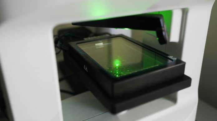
EPFL scientists have recently come up with a next-generation microscope that can show live cells function and react under various experimental conditions. Called as CX-A, the microscope will show scientists how their organelles interact and react to stimuli.
This discovery could pave the way significant revelations about biological procedures that until now have been inadequately comprehended in view of the absence of a solid method to observe them.
With a resolution of <200 nm, scientists can see living cell populations and zoom in all the way down to individual organelles.
Samples are prepared by placing the cells on a special 96 well plate. Scientists can quickly set up their examinations by essentially indicating how frequently they need pictures to be taken; the device at that point keeps running without anyone else.
Information can be gathered along these lines for whatever length of time that required, with a huge number of images assumed taken over a time of a few days or weeks. Therefore, they can get remarkable bits of knowledge into how natural procedures work, how organelles collaborate, and how mitochondria form intricate networks, for instance.
Originally, Nanolive’s CEO Yann Cotte first developed this technology, when he was a Ph.D. student. The technology works like an MRI machine that generates images of cells from all angles using their refractive index and then compiles 3D images with the help of advanced software.
A rotating laser illuminates the sample at a 45° angle to create a hologram, giving a novel investigation cell under natural conditions. The technique is non-invasive, manipulation free, and obstruction-free, and the rotational scanning takes into account 3D reproduction with excellent resolution.
Unlike traditional microscopes, Nanolive technology requires no stains.
Mathieu Frechin, head of quantitative biology at Nanolive said, “With our microscope, scientists can run experiments under a range of conditions and obtain high-quality images without adding fluorescent markers. The images generated using the refractive index and the possibility to combine them with fluorescent signals enable scientists to follow over time dynamic and delicate cell processes – such as the membrane potential – of living subcellular structures like mitochondria. The signals reveal subtle variations in structure and activity that occur in response to drugs or genetic mutations.”
Lisa Pollaro, chief marketing officer said, “We plan to double our headcount by next year so we urgently need to move into bigger offices. Besides that, we have plenty of research projects in the pipeline, although I can’t discuss them in detail. All I can say is that new breakthroughs are coming.”
