
The mind-boggling and secretive mechanisms that drive correspondence and responses inside human cells could be very nearly being unwound because of a spearheading new method.
Specialists from the Universities of Leeds, Exeter, and Cambridge have tackled an imaginative new technique to pick up a more noteworthy comprehension of flagging stations inside the phones, called nanodomains.
They trust the new method could prepare for a more noteworthy comprehension of the reasons for conceivably perilous conditions, for example, coronary illness, and in addition, potential new treatment pathways.
The nanodomains are known to drive crucial physiological procedures in the body, including the beginning of malady.
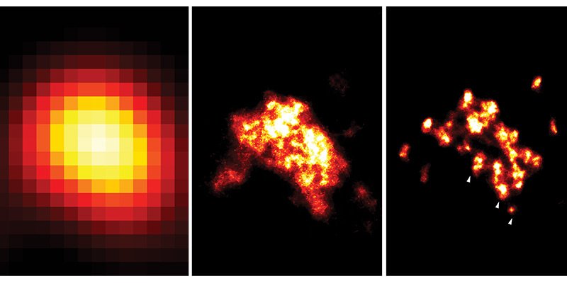
Researchers have, as of not long ago, for the most part, depended on electron-microscopy to examine these structures. However, the innovation has not enabled access to better instruments of the nanodomains at a sub-atomic level.
Presently, the UK examine group has refined another light-based super-determination microscopy strategy that permits top-notch imaging of the flagging stations in the human heart.
First author Dr. Isuru Jayasinghe said, “This new super-resolution microscopy tool gives us the perfect window to visually examine the individual protein changes within heart cells’ molecular machinery which lead to heart failure.”
“At present, none of the treatments or therapies provided to heart failure patients specifically target the signaling stations – nanodomains – within the cell, which the evidence overwhelmingly suggests are a major cause of heart disease.”
“We believe that by visualizing these signaling structures at this level of detail using super-resolution microscopy we can help guide investigations into how we can target or repair these molecular machines and thus, in the long term, help patients to overcome heart disease.”
Aptitude in the outline of engineered DNA strands was given by Dr. Lorenzo Di Michele from the University of Cambridge.
The pivotal new procedure enables researchers to stick point any number of particular kinds of proteins inside the cells, the checking of every species of protein, and perceptions of the exact examples in which they are masterminded.
Professor Christian Soeller, who led the study said, “Slightly more than a decade ago nobody thought that we would ever see individual molecules with light, the resolution just seemed insufficient to resolve such fine detail.”
“Since then an astonishing array of new tricks has been devised. In our latest advance, the use of synthetic DNA has been critical – the deep understanding of the chemistry of DNA we have today makes it an enormously versatile tool.”
Accordingly, the group says that their examination gives a “flawless window” to inspect the progressions that happen in the sub-atomic apparatus, which are a noteworthy reason for heart disappointment.
They trust the additional visual detail that the new imaging gives will manage more definitive examinations concerning how to target or repair these flagging stations or the atomic machines inside them all the more decisively.
The research paper ‘True Molecular Scale Visualization of Variable Clustering Properties of Ryanodine Receptors‘ is published 9 January 2018 in the journal Cell Reports.