
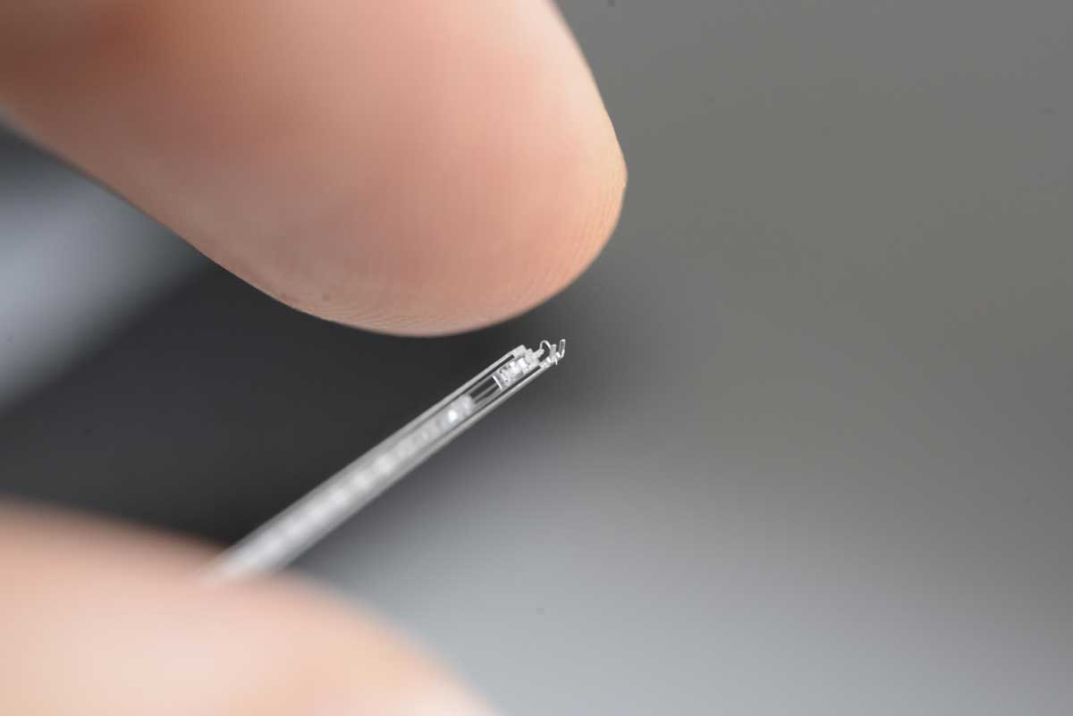
On September 11, a team of scientists has presented a breakthrough device at EPFL- called SPOT-RVC that doctors could use to inject medicine into retinal veins with unprecedented accuracy. The device is originally a microscopic glass with a tiny fluidic channel no wider than a strand of hair as well as a sophisticated mechanism of flexible blades.
The SPOT-RVC device which stands for Safe Puncture Optimized Tool for Retinal Vein Cannulation is just 6cm in length and 1 mm in thickness. Scientists developed this device to inject medicine directly into a patient’s retinal veins – something that has never before been possible.
In addition, it has a tiny fluidic channel, narrow than a strand of hair and also has a sophisticated mechanism of flexible blades. The channel was developed using an innovative process developed by scientists at Galatea, that allows for fabricating, arbitrarily long and shaped, sealed cavities. Interestingly, the channel extends all the way down to the needle tip, thus allow doctors to inject the medicine.
Finally, the device is made of a single piece of fused silica (SiO2), with the help of unique expertise of FEMTOprint for integrating multiple functions in the same substrate.
Professor Thomas J. Wolfensberger, the chief physician at Jules-Gonin Hospital, said, “We wanted to develop a surgical method for treating retinal vein occlusion, which occurs when the main vein carrying blood away from the eye is blocked. There is currently no way to treat this condition – we can only treat the resulting complications.”
“And those complications can be severe. When a blood clot blocks the retinal vein, this reduces the amount of oxygen carried to the retina and can trigger sudden vision loss. Over 16 million people around the world suffer from this condition, which mostly afflicts the elderly.”
As veins are so small, it’s quite challenging to get the needle into the vein without over puncturing. It’s like if you want to drill a hole into a plank of wood but don’t want the hole to go all the way through.
But, the device seems to solve that problem also: using the device, scientists could inject blood-clot-dissolving compounds directly into patients’ retinal veins safely, without damaging the surrounding tissue.
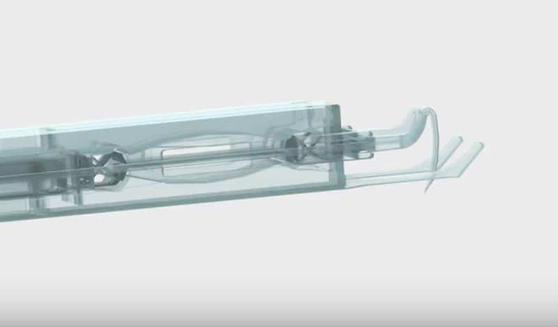
Dr. Charles Baur, a senior scientist at Instant-Lab, said, “We, drew on Instant-Lab’s expertise in flexible microstructures and multistable systems to engineering a microscopic device (< 1 mm in diameter) that can transition from one stable state to another very quickly – in around a millisecond – and a controlled manner. With our dynamic perforation mechanism that controls both the penetration force and direction of the needle, retinal veins don’t have time to deform. Also, the penetration force is independent of the force exerted by the surgeon’s hand, which limits the risk of over puncturing.”
Dr. Baur said, “Since it’s monolithic, there’s no assembly required – a step that would be nearly impossible and would make it very difficult to sterilize the instrument.”
The complex monolithic integration with high precision was made possible because of FEMTOprint. FEMTOprint uses ultrafast lasers 3D printing and proprietary post-processing techniques. What’s more, the expertise from the Galatea lab helps to understand ultrafast laser-matter interactions and its use for making complex micro-devices, such as optofluidics and optomechanical devices.
FEMTOprint presented SPOT-RVC at the Swiss high-precision industry convention (EPHJ), which was held this past June in Geneva. The device won the 2019 Exhibitors’ Grand Prix – an encouraging start.
“We got good results from our in vitro and in vivo tests. Now we need to conduct preclinical trials and obtain the necessary certifications. Then we’ll move on to the production stage, which will require a fairly large investment. I genuinely hope that one day, the device will become a useful tool for eye surgeons.”
The device was developed through a joint R&D project involving two EPFL Neuchâtel labs (Instant-Lab and Galatea), the Jules-Gonin Hospital of Ophthalmology in Lausanne and Ticino-based FEMTOprint.