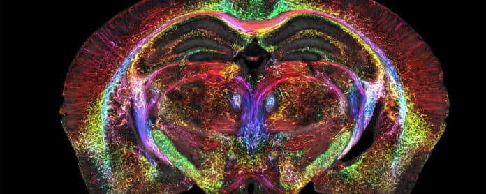
MRI eases the visualization of soft, watery tissue that is difficult to detect with X-rays. Even if MRI offers good resolution to spot a brain tumor, it still needs to be much sharper to visualize microscopic details within the brain that reveal its organization.
Researchers took up the challenge and improved the resolution of MRI, leading to the sharpest images ever captured of a mouse brain in a decades-long technical feat led by Duke‘s Centre for In Vivo Microscopy with colleagues at the University of Tennessee Health Science Centre, the University of Pennsylvania, University of Pittsburgh, and Indiana University.
Scientists created mouse brain images that are significantly crisper than a standard clinical MRI for humans to commemorate the 50th anniversary of the first MRI. This is the scientific equivalent of going from an 8-bit pixelated image to the hyper-realistic detail of a Chuck Close painting.
The new images’ voxels, or what you may refer to as cubic pixels, are only 5 microns in size. Compared to a clinical MRI voxel, it is 64 million times smaller.
Researchers mainly focused their magnets on mice. The refined MRI offers a significantly new way to visualize the connectivity of the entire brain at record-breaking resolution. These new insights from mouse imaging could pave the way toward a better understanding of human conditions, such as how the brain changes with age, diet, or even with neurodegenerative diseases like Alzheimer’s.
G. Allan Johnson, Ph.D., the lead author of the new paper and the Charles E. Putman University Distinguished professor of radiology, physics, and biomedical engineering at Duke, said, “It is truly enabling. We can start looking at neurodegenerative diseases in an entirely different way.”
Some of the essential components include a powerful magnet (the majority of clinical MRIs use magnets between 1.5 and 3 Tesla; Researchers use a 9.4 Tesla magnet), a unique set of gradient coils that are 100 times more powerful than those in a clinical MRI and aid in creating the brain image, and a high-performance computer that is roughly equivalent to nearly 800 laptops working simultaneously to image one brain.
After “scanning the daylights out of it,” Johnson and his team send the tissue over to be scanned with a different method called light sheet microscopy. They can label particular populations of brain cells with this supplementary method, such as dopamine-producing cells, to track the progression of Parkinson’s disease.
The team then overlays the original MRI scan, which is far more anatomically precise and offers a vivid view of cells and circuits throughout the entire brain, with the light sheet images, which provide a highly accurate look at brain cells.
Researchers may now look into the tiniest mysteries of the brain in ways that have never been conceivable before, thanks to this unified whole-brain data imaging.
Thanks to this whole brain data imagery, researchers can now peer into the microscopic mysteries of the brain in ways never possible before.
One set of MRI images shows how brain-wide connectivity changes as mice age and how specific regions, like the memory-involved subiculum, change more than the rest of the mouse’s brain.
Another set of images showcases a spool of rainbow-colored brain connections that highlight the remarkable deterioration of neural networks in a mouse model of Alzheimer’s disease.
The hope is that by making the MRI an even higher-powered microscope, Johnson and others can better understand mouse models of human diseases, such as Huntington’s disease, Alzheimer’s, and others. And that should lead to a better understanding of how similar things function or go awry in people.
Johnson said, “Research supported by the National Institute of Aging uncovered that modest dietary and drug interventions could lead to animals living 25% longer. So, the question is, is their brain still intact during this extended lifespan? Could they still do crossword puzzles? Will they be able to do Sudoku even though they’re living 25% longer? And we have the capacity now to look at it. And as we do so, we can translate that directly into the human condition.”
Journal Reference:
- G. Allan Johnson, Yuqi Tia, David G. Ashrook. Merged magnetic resonance and light sheet microscopy of the whole mouse brain. PNAS. DOI: 10.1073/pnas.2218617120
