
While crystallization is a ubiquitous and necessary process, the microscopic picture of crystal nucleation is yet to be established. It is important to develop the tiny picture of crystal nucleation for both academic and industrial research.
Scientists at the University of Geneva (UNIGE) reported successful visualization of crystal nucleation. They used single-crystal nucleation spectroscopy (SCNS) combined with Raman microspectroscopy and optical trapping induced crystallization to investigate one crystal nucleation at a time.
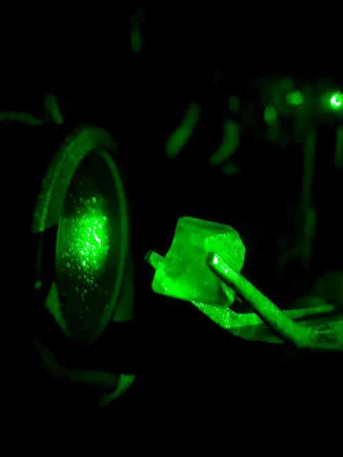
Oscar Urquidi, a doctoral student in the Department of Physical Chemistry and co-first author of this research, said, “We used lasers to highlight the molecular structure during the nucleation but also to induce the nucleation phenomenon and thus be able to observe it and record its spectral imprint. The model substance chosen to conduct these experiments was glycine, an amino acid that is an essential building block of life, dissolved in water.”
Johanna Brizard, a researcher in the Department of Physical Chemistry and co-first author of the research, said, “We have succeeded in demonstrating and visualizing the organization and formation of molecular aggregates that precede crystallization.”
Takuji Adachi, assistant professor in the Department of Physical Chemistry at the UNIGE Faculty of Science, said, “Our work has revealed a stage of crystallization that was previously invisible. Visualizing more precisely and better understanding what is happening at the molecular level is useful for directing certain manipulations more effectively. In particular, this discovery could make it easier to obtain purer and more stable crystal structures for certain substances used in the design of many drugs or materials.”
Journal Reference:
- Oscar Urquidi, Johanna Brazard et al. In situ optical spectroscopy of crystallization: One crystal nucleation at a time. DOI: 10.1073/pnas.2122990119