
Cancer is the fifth leading cause of death in India and second leading in the USA, but it can be preventable if diagnosed early. Patients have to face many biopsies, and too many tests run on the body, making them more time-consuming procedures. Now, researchers from Japan are working on developing new techniques that make cancer testing much less invasive.
Tokyo Medical and Dental University researchers discover ion-sensitive field-effect transistors. Combined with enzymatic chemical signal amplification, these transistors help detect cancer-related makers in breast cancer cells. The study was published in September in the Journal of the American Chemical Society; researchers from Tokyo Medical and Dental University have disclosed a new technique for detecting cancer cell markers. This technique is a dynamic way to determine the diagnosis, prognosis, and successful treatment. This modern technique can detect cancer markers in a non-invasive way to monitor and evaluate patients.
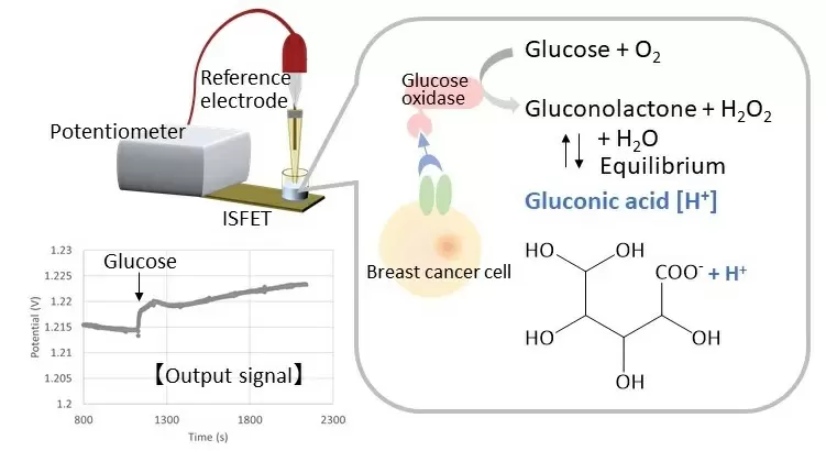
In this technique, epidermal growth factor receptor (EGFR) detection using a cell-based field-effect transistor (FET) with enzymatic chemical signal amplification is put forward. Here four human breast cancer cell lines [BT474, MDA-MB-231(MM231), MDA-MB-231(MM231), MDA-MB-468(MM468), and MDA-MB-453(MM453)] were used to compare the expression levels of RGFR.
The cells were non-specifically captured on the surface of the gate of the FET, with consideration of their surface antigens. With this arrangement, the heterogeneity of the cells was analyzed using secondary antibodies, which were then combined with different types of enzymes. Then four breast cancer cell lines with different levels of EGFR expression were shown on the surface of the extracellular matrix (ECM) gel-coated gates of the FETs. Glucose oxidase (Gox) was combined with the secondary antibody, and output signals of the cell-based FETs changed, depending on expression levels of EGFR upon the addition of glucose.
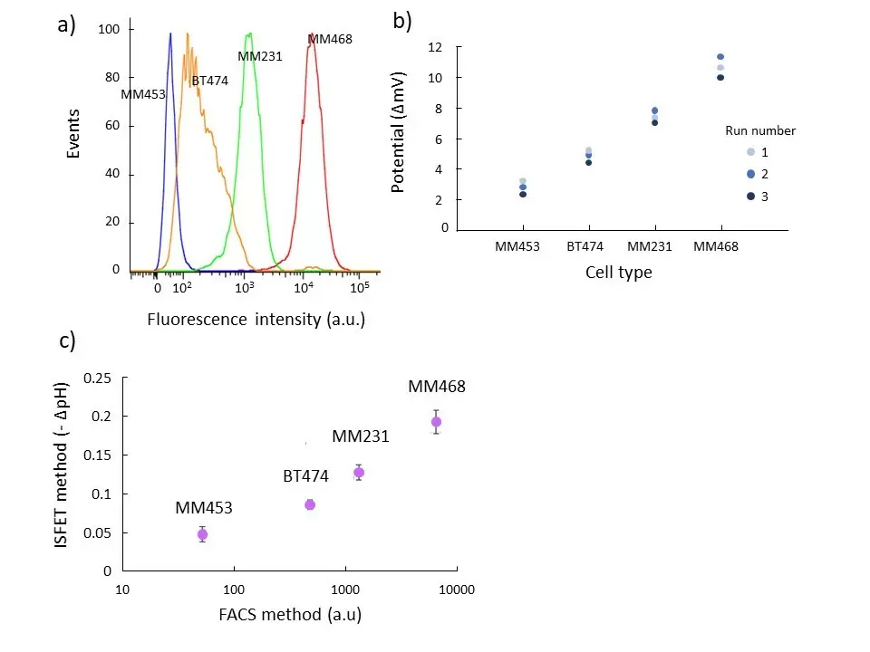
The order of expression levels of EGFR among the four cell lines, considered with the cell-based FETs, was consistent with the result of fluorescence detection determined by fluorescence-activated cell sorting (FACS). These cell-based FETs are useful for miniaturization and analyses of target molecules expressed on the membranes of cells and EVs. Size and cost-effectiveness for cancer testing could authorize their understanding of a future liquid biopsy.
The first author of the study Miyuki Tabata, states, “Circulating tumor cell s(CTCs) which are cancer cells found in blood are main targets used to evaluate cancer patients blood samples,”
“However, it can be challenging to differentiate these cells from the blood, and current approaches do not adequately detect both epithelial cell and mesenchymal cell markers, which are important for determining the stage of cancer.”
So, to have a quick and easy detection of cancer-related markers on CTCs, researchers used an apparatus called an ion-sensitive field effect transistor which is a tiny electrical circuit that is activated by a change in pH. These transistors were coated with breast cancer cells with an antibody linked to a chemical reporter that causes a change in pH if the antibody recognizes the cells.
Yuji and Miyahara, the senior author, found that “the chemical reporter glucose oxidase successfully detected the expression of epidermal growth factor receptor (EGFR) a marker of poor cancer prognosis, on CTC membranes”.
“Furthermore, the strength of the chemical signal correlated with the amount of EGFR expressed by the cells.”
“These results provide proof of concept that an ISFET-based system can be used to efficiently assess patient cancer status based on liquid biopsy samples,” states Tabata.
This technique has the capability to authorize high-throughput analysis of cancer cells at single-cell resolution. Even using multiple antibodies for the chemical enzyme detection procedure could enable the analysis of numerous cancer-related markers.
This procedure is the more easy investigative procedure for doctors and other researchers as it is a non-invasive technique and easy to detect cancer cell markers.
Journal Reference
- Miyuki Tabata, Xinyue Liu, Chattarika Khamhanglit, Sayo Kotaki, and Yuji Miyahara. Detection of Epidermal Growth Factor Receptor Expression in Breast Cancer Cell Lines Using an Ion-Sensitive Field-Effect Transistor in Combination with Enzymatic Chemical Signal Amplification. American Chemical Society 2022, 144, 36, 16545 DOI: 10.1021/jacs.2c06122