
Acquiring data from almost 300 neurosurgical patients who have implanted electrodes directly on the brain, scientists at the University of Pennsylvania have constructed the first whole-brain map of electrical connectivity. They demonstrated that the low-frequency rhythms that drive communication between the frontal, temporal and medial temporal lobes, key brain regions that engage during memory processing.
Many previous studies examined brain networks using non-invasive tools. But the observations at large-scale networks using direct human-brain recordings have been difficult to secure because these data can only come from neurosurgical patients. The study illustrates the way unique locales of the mind convey amid subjective procedures like memory arrangement.
Scientists acquired data from multiple hospitals across the country. They involved patients who underwent clinical monitoring for seizures. Later, they asked patients to perform a free-recall memory task that asked them to view a series of words on a screen, then repeat back as many as they could remember.
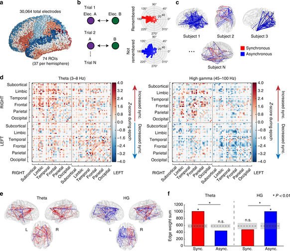
During this, they also observed brain activity occurring on slow and fast time scales. They found that when a man is viably making new recollections – for this situation, recalling that one of the introduced words – an arrangement between mind areas has a tendency to reinforce with moderate floods of action yet debilitate at higher frequencies.
Solomon, the paper’s lead author said, “We found that the low-frequency connectivity of a brain region was associated with increased neural activity at that site.”
“This suggests that, for someone to form new memories, two functions must happen simultaneously: brain regions must individually process a stimulus, and then those regions must communicate with each other at low frequencies.”
Michael Kahana, Penn professor of psychology said, “Better understanding the brain networks that activate during memory processing. This gives us a better ability to fine-tune electrical stimulation that might improve it.”
“We’re now prepared to ask whether we can use measures of functional connectivity to guide our choice of which brain region to target with electrical stimulation. Ultimately, given the size of this dataset, these discoveries would not be possible without years of effort on the part of our participants, clinical teams, and research scientists.”
Now scientists are planning to examine the interaction between brain stimulation and the functional connections that the study uncovered.