Ultrasound has advantages compared to other imaging methods, including being nonionizing, relatively low cost, and portable. Current embodiments of ultrasound technology range from cart-based bedside systems to portable hand-held devices.
The method requires the placement of piezoelectric transducers in contact with the patient to transmit and then detect reflected and scattered acoustic waves at the body surface. This case may be limiting in situations where clinicians might want to image patients who don’t tolerate the probe well, such as babies, burn victims, or other patients with sensitive skin.
Now, MIT scientists have found a solution to this; they have developed a new laser ultrasound technique, an alternative to ultrasound that doesn’t require contact with the body to see inside a patient. The method leverages an eye- and a skin-safe laser system to remotely image the inside of a person.
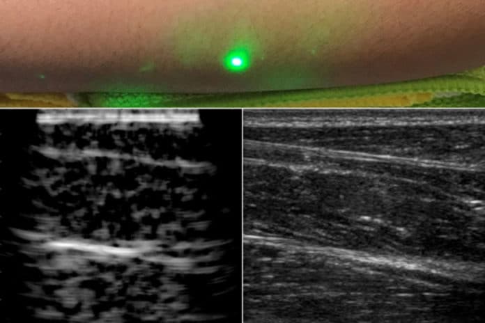
For testing, scientists imaged the forearms of several volunteers and observed common tissue features such as muscle, fat, and bone, down to about 6 centimeters below the skin. These images, comparable to conventional ultrasound, were produced using remote lasers focused on a volunteer from half a meter away.
Brian W. Anthony, a principal research scientist in MIT’s Department of Mechanical Engineering and Institute for Medical Engineering and Science (IMES), said, “We’re at the beginning of what we could do with laser ultrasound. Imagine we get to a point where we can do everything ultrasound can do now but at a distance. This gives you a whole new way of seeing organs inside the body and determining properties of deep tissue, without making contact with the patient.”
To develop this technique, scientists selected 1,550-nanometer lasers, a wavelength that is highly absorbed by water (and is eye- and skin-safe with a large safety margin).
Scientists tested this idea with a laser setup, using one pulsed laser set at 1,550 nanometers to generate sound waves, and a second continuous laser, tuned to the same wavelength, to remotely detect reflected sound waves. This second layer is a sensitive motion detector that measures vibrations on the skin surface caused by the sound waves bouncing off muscle, fat, and other tissues. Skin surface motion, generated by the reflected sound waves, causes a change in the laser’s frequency, which can be measured. By mechanically scanning the lasers over the body, scientists can acquire data at different locations and generate an image of the region.
Scientists are now planning to improve their technique, and they are looking for ways to boost the system’s performance to resolve fine features in the tissue. They are also looking to hone the detection laser’s capabilities. Further down the road, they hope to miniaturize the laser setup, so that laser ultrasound might one day be deployed as a portable device.
This research was supported in part by the MIT Lincoln Laboratory Biomedical Line Program for the United States Air Force and by the U.S. Army Medical Research and Material Command’s Military Operational Medicine Research Program.
The paper is published in Nature in the journal Light: Science and Applications.
