
Scientists at the Universities of Canterbury and Otago have developed a revolutionary new 3D color medical scanner that is expected to revolutionize medical imaging globally. Dubbed as MARS, the scanner offers far greater detail of the body’s chemical components.
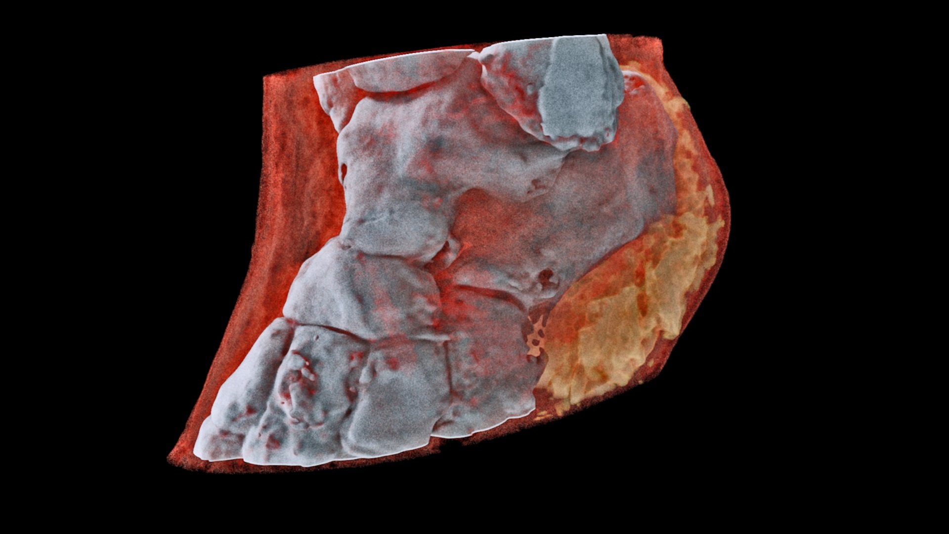
The MARS spectral X-ray scanner produces images with significantly improved diagnostic information. It measures the X-ray spectrum to produce color images instead of black-and-white ones and shows different components of body parts such as fat, water, calcium, and disease markers.
In order to develop this scanner, scientists used technology used by the European Organization for Nuclear Research (CERN) in the hunt for the ‘God particle’ in a medical scanner.
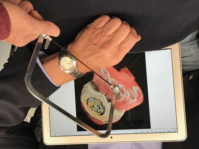
Small versions of the scanner that can house tissue samples are, as of now, being used in research about foundations around the globe. The first human has now been scanned over a larger form of the scanner. Professor Phil Butler was the main individual to be checked. His lower leg and wrist were imaged.

The next step in development is an imminent clinical trial where orthopedic and rheumatology patients from Christchurch will be scanned. This will allow the MARS team to compare the images produced by their scanner with the technology currently used in New Zealand hospitals.
Professor Anthony Butler says, “after a decade in development, it is really exciting to have reached a point where it’s clear the technology could be used for routine patient care.”
“X-ray spectral information allows health professionals to measure the different components of body parts such as fat, water, calcium, and disease markers. Traditional black-and-white x-rays only allow measurement of the density and shape of an object.”
“So far, researchers have been using a small version of the MARS scanner to study cancer, bone and joint health, and vascular diseases that cause heart attacks and strokes. In all of these studies, promising early results suggest that when spectral imaging is routinely used in clinics, it will enable more accurate diagnosis and personalization of treatment.”