Anxiety is normal and critical to an animal’s safety. Anxiety is an emotional response to a distant threat—being in an environment that exposes an animal to predators, for example. The safe bet is to sidestep those environments, so anxiety kicks in avoidance behaviors.
When people overestimate threats—when talking to a crowd invokes the same response as potentially running into a snake—anxiety becomes a problem.
A new study by the scientists at Columbia University Irving Medical Center (CUIMC) has identified anxiety” cells deep inside your brain.
The scientists found the phones in the brains of mice, inside a structure called the hippocampus. Yet, the cells likely likewise exist in people, says Rene Hen, Ph.D., educator of neuroscience and pharmacology (in psychiatry) at Columbia and one of the examination’s senior specialists.
“We call these tension cells since they just fire when the creatures are in places that are inherently alarming to them,” Hen says. “For a mouse, that is an open zone where they’re more presented to predators or a lifted stage.”
The terminating of the uneasiness cells sends messages to different parts of the cerebrum that turn on edge practices; in mice, those incorporate maintaining a strategic distance from the unsafe region or escaping to a sheltered zone.
In spite of the fact that numerous different cells in the cerebrum have been recognized as assuming a part in nervousness, the cells found in this examination are the primary known to speak to the condition of tension, paying little mind to the sort of condition that incites the feeling.
“This is energizing since it speaks to an immediate, quick pathway in the mind that gives creatures a chance to react to uneasiness inciting places without expecting to experience higher-arrange cerebrum locales,” says Mazen Kheirbek, Ph.D., associate educator of psychiatry at UCSF and the examination’s other senior examiner.
“Since we’ve discovered these phones in the hippocampus, it opens up new regions for investigating treatment thoughts that we didn’t know existed earlier,” says the examination’s lead creator Jessica Jimenez, Ph.D., an MD/ Ph.D. understudy at Columbia University’s Vagelos College of Physicians and Surgeons.
The discoveries show up in the Jan. 31 online issue of Neuron.
“This examination demonstrates how translational research utilizing essential science procedures in creature models can explain the basic premise of human feelings and purposes behind a mental disorder, in this way pointing the path for treatment improvement,” says Jeffrey Lieberman, MD, the Lawrence C. Kolb Professor and Chair of Psychiatry at Columbia.
To understand how things go awry in anxiety disorders, researchers in the Hen lab have been looking at mice to decipher how the brain processes healthy anxiety.
Dr. Kheirbek, who was an assistant professor at CUIMC before moving to UCSF said, “We wanted to understand where the emotional information that goes into the feeling of anxiety is encoded within the brain.”
The hippocampus assumes an outstanding part of the mind’s capacity to frame new recollections and to enable creatures—from mice to people—to explore complex conditions. Be that as it may, late research has likewise ensnared the hippocampus in managing temperament, and studies have demonstrated adjusting cerebrum movement in the central piece of the hippocampus can diminish uneasiness. It’s likewise realized that the hippocampus sends signs to different regions of the cerebrum—the amygdala and the hypothalamus—that have additionally been appeared to control nervousness related conduct.
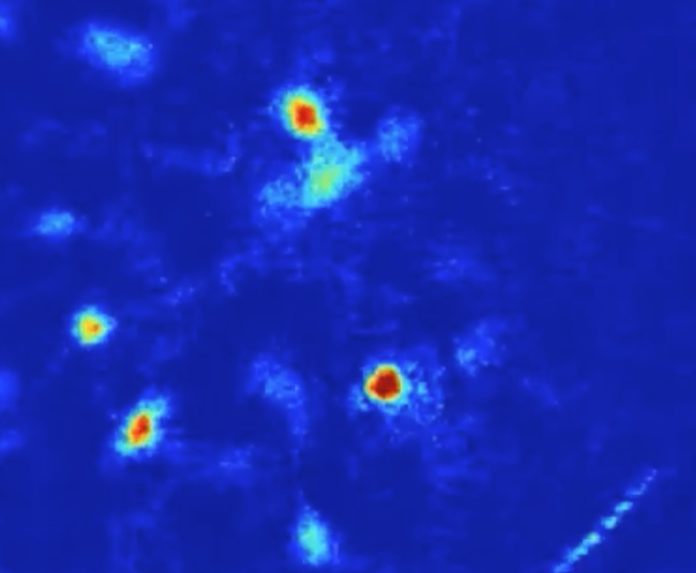
Scientists used a miniature microscope to record the activity of hundreds of cells in the hippocampus in the mice s the mice freely moved around their surroundings.
At whatever point the creatures were in uncovered, tension inciting situations, the analysts saw that particular cells in the ventral piece of the hippocampus were dynamic. What’s more, the more on edge the mice appeared, the more prominent the movement in the cells.
The analysts followed the yield of those cells to the hypothalamus, which is known to control practices related with uneasiness (in individuals, those incorporate expanded heart rate, evasion, and emission of stress hormones).
By killing the tension cells and on utilizing a method called optogenetics that enables researchers to control the action of neurons utilizing light emissions, the scientists found that the uneasiness cells control nervousness practices. At the point when the phones were hushed, the mice quit delivering dread related practices, meandering onto lifted stages and far from defensive dividers. At the point when the uneasiness cells were animated, the mice showed more dread practices notwithstanding when they were in “safe” environment.
The disclosure of the nervousness cells raises the likelihood of discovering medications that objective them and lessen uneasiness. “We’re hoping to check whether these cells are distinctive molecularly from different neurons,” Hen says. “On the off chance that there’s a particular receptor on the cells that recognize them from their neighbors, it might be conceivable to create another medication to diminish tension.”
