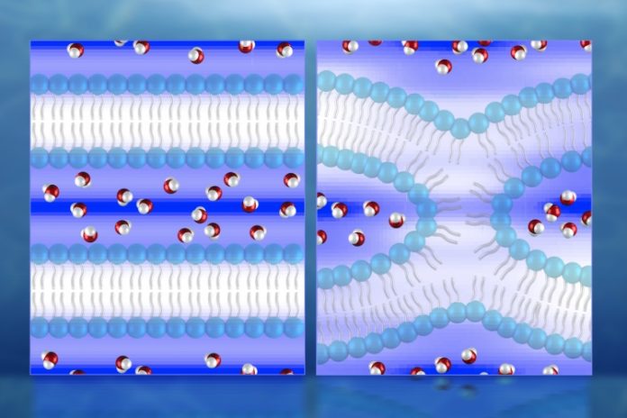
New 3D maps of water distribution amid cellular membrane combination are quickening logical comprehension of cell development, which could prompt new medications for infections related to cell fusion. Utilizing neutron diffraction at the Department of Energy’s Oak Ridge National Laboratory, scientists have made the principal coordinate observations of water in lipid bilayers used to show cell membrane fusion.
The exploration could give new bits of knowledge into diseases in which normal cell fusion is disturbed, for example, Albers-Schönberg disease (osteopetrosis), help encourage the advancement of fusion-based cell therapies for degenerative diseases, and prompt medicines that forestall cell-to-cell fusion between cancer cells and non-cancer cells.
At the point when two cells combine amid fertilization, or a membrane-bound vesicle fuses amid viral section, neuron signaling, placental development, and numerous other physiological capacities, the semi-porous film bilayers between the fusing partners must be converged to trade their inward contents.
As the two membranes approach one another, hydration forces increment exponentially, which requires a lot of vitality for the membranes to survive. Mapping the distribution of water particles is vital to understanding the fusion procedure.
Scientists used the small-angle neutron scattering (EQ-SANS) instrument at ORNL’s Spallation Neutron Source and the biological small-angle neutron scattering (Bio-SANS) instrument at the High Flux Isotope Reactor, both of which can probe structures as small as a few nanometers in size.
Durgesh K. Rai, co-author said, “We used neutrons to probe our samples, because water typically can’t be seen by x-rays, and because other imaging techniques can’t accurately capture the extremely rapid and dynamic process of cellular fusion. Additionally, the cold, lower-energy neutrons at EQ-SANS and Bio-SANS won’t cause radiation damage or introduce radicals that can interfere with lipid chemistry, as x-rays can do.”
The outcoming water density map revealed that the water separates from the lipid surfaces in the underlying lamellar, or layered, stage. In the intermediate fusion stage, known as hemifusion, the water is altogether diminished and squished into pockets around a stalk—a exceptionally curved lipid “bridge” associating two membranes before fusion completely happens.
Shuo Qian, the study’s corresponding author and a neutron scattering scientist at ORNL said, “For the neutron scattering experiments, we replaced some of the water’s hydrogen atoms with deuterium atoms, which helped the neutrons observe the water molecules during membrane fusion. The information we obtained could help in future studies of membrane-acting drugs, membrane-associated proteins, and peptides in a membrane complex.”
The study is published in the Journal of Physical Chemistry Letters.
