
How genes control the formation of entire organisms or morphogenesis of an organ? This is the most fundamental question in biology.
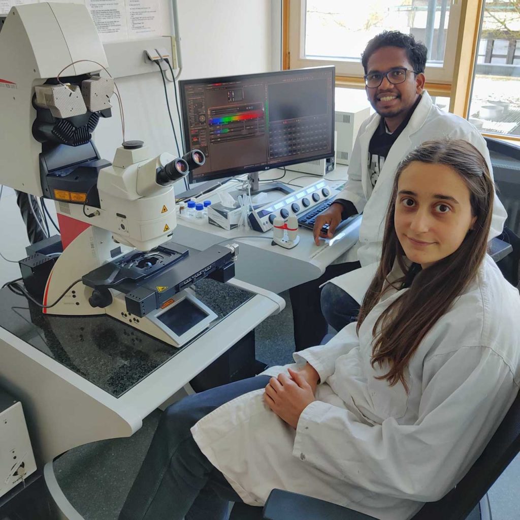
The term morphogenesis means an intricate process that incorporates many processes that work on various scales – from the molecular control of gene expression to cell coordination in a tissue. To better represent the morphogenesis, an interdisciplinary team of biologists, physicists, and computer scientists from several scientific institutions set up the digital image analysis pipeline “PlantSeg,” based on machine learning.
Scientists from the TUM School of sciences, for the first time, have determined the cellular composition of the ovules of the model plant Arabidopsis thaliana using the latest microscopy methods with image processing based on machine learning. For the first time, scientists made it possible to display an organ’s complete development series using digital 3D ovules.
Using plants and state of the art microscopy, scientists created a digital 3D atlas of the development of ovules of Arabidopsis thaliana. The atlas is characterized by complete cellular dissolution, even in deep tissue layers.
It enables quantitative spatiotemporal analysis of cellular and gene expression patterns with cell and tissue resolution. For example, the unusual establishment of a new tissue layer in the ovule could be described for the first time, and specific cell division patterns in deeper layers could be associated with the ovule’s curvature.
Scientists noted, “the cellular distribution of the gene expression of an important regulatory gene could be described quantitatively. These results can now lay the foundation for the next generation of studies on morphogenesis.”
The work was carried out as part of the research group FOR 2581 “Plant Morphodynamics” funded by the German Research Foundation. The research group includes TUM, EMBL Heidelberg, the Max Planck Institute for Breeding Research in Cologne, Heidelberg University, and the John Innes Center in Norwich, UK. The research group members consist of biologists, physicists, and computer scientists whose common interest lies in understanding how plants shape their organs.
Journal Reference:
- Athul Vijayan et al. A digital 3D reference atlas reveals cellular growth patterns shaping the Arabidopsis ovule. DOI: 10.7554/eLife.63262