
The cornea, a focal point holed up behind the pupils, focuses light from the screen to the back of the eye. Be that as it may, if the cornea’s horizontal and vertical centering power is unique, the vision will be blurred. For example, the vertical and even lines which frame the letter “T” won’t be united in the center.
To avoid this centering imperfection, an artificial lens is ideally designed to change the state of the light wavefronts from planar into perfectly round, as it is believed that spherical wavefronts essentially focus at their unique center of curvature.
An international team of researchers, affiliated with UNIST have discovered a new property of wave propagation that leads to an all-new way to improve the resolution of virtually all optical technologies, including microscope lenses, telecommunications, laser-based lithography, and biological and astronomical imaging.
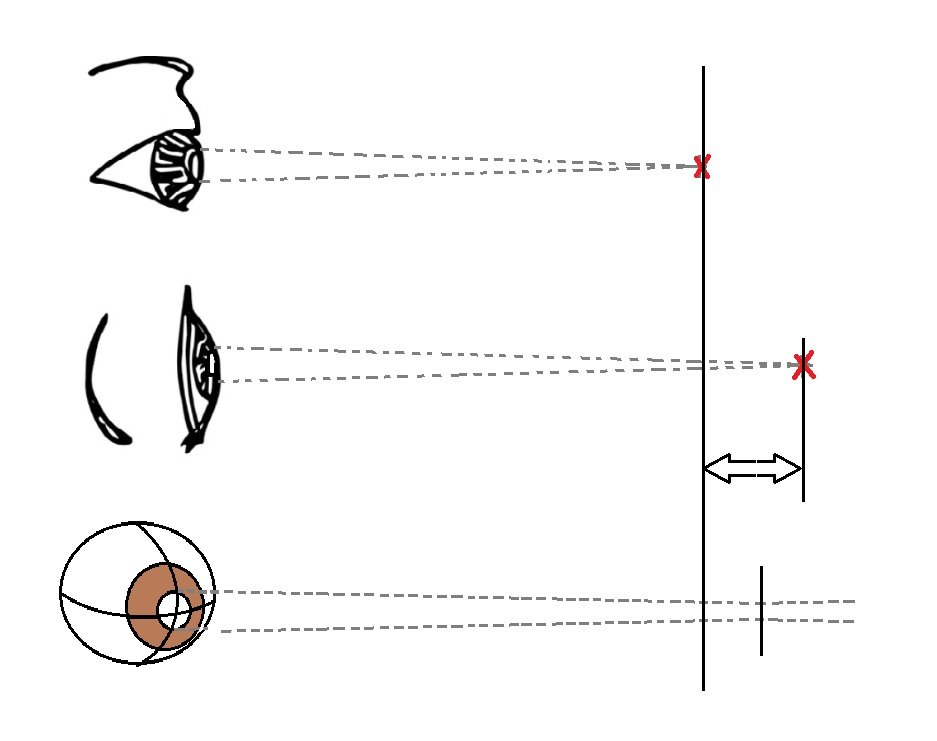
Professor François Amblard in School of Life Sciences and his research team at the Center for Soft and Living Matter said, “A full spherical wave is symmetric and has its focus exactly at the center of the sphere. However, to keep this spherical symmetry, the light should propagate in all directions onto the sample. And this virtually never happens.”
“In the case of the pond, this would be similar to going back in time to try to refocus a limited wave arc, instead of the full circle: they would not necessarily converge into the same impact point, meaning that information about the center location is lost. Practically, wavefronts are passed through an aperture that is limited to a portion of a sphere. Consequently, spherical symmetry is broken and the information is lost.”
Scientists explained, as the aperture gets smaller, the focus moves increasingly in reverse towards the lens, with the end goal that the underlying concentration is never again “in focus.” As a result, if the aperture isn’t equivalent in the vertical and horizontal planes, central movements will vary between these bearings, prompting astigmatism.
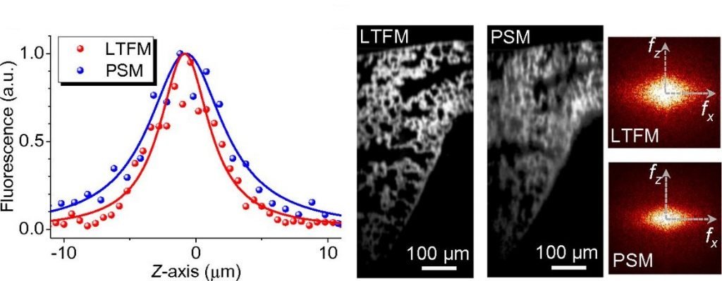
The team applied the idea to improve a technique called line-temporal focusing microscopy (LTFM, also named spatiotemporal focusing), which makes use of a naturally asymmetric input beam. As LTFM is a method used to visualize deep biological structures, the researchers tested their focal shift correction strategy with mouse lung tissues. An unprecedented resolution was obtained that even outperformed a classical technique called point scanning microscopy (PSM).
Even though this effect is very small and can be neglected for ordinary applications, correcting for aperture-induced astigmatism could make a significant difference in delicate systems, like advanced microscopy used to acquire big date. Understanding that astigmatism is intrinsic to the broken circular symmetry could help design corrections tailored to the aperture shape, especially in fields such as astronomy, telecommunication, or with ultrasounds, where non-circular apertures cannot be avoided.
Professor Steve Granick, co-correspondent author of this study said, “In the future, we plan to apply aperture-induced astigmatism to even more complex information transfer technologies. Moreover, the study opens avenues to basically improve the design of any equipment handling electromagnetic waves, ultrasounds, or particles beams. For example, it also applies to waves, used with space antennas to focus on a satellite or spaceship. We believe it can contribute to design better systems in synthetic microscopic eyesight, telecommunications, and even microwave devices.”
The findings of this research have been published in Proceedings of the National Academy of Sciences of the United States of America (PNAS) on June 13, 2018.