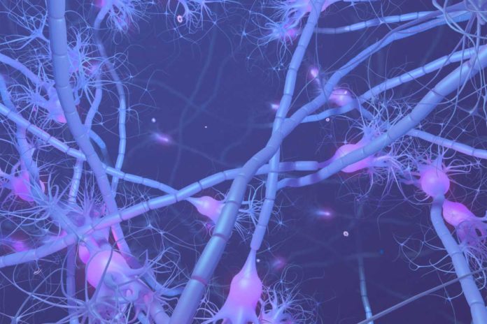
In a new study by the Harvard’s Department of Stem Cell and Regenerative Biology in collaboration with a joint department between Harvard Medical School and the Faculty of Arts and Sciences, scientists discovered that parts of the neuron are far more mind-boggling than once thought.
This study adds another twist in the ever-evolving understanding of the nerve cells that make up our brains. Neurons are the cells that make up the brain and the nervous system. They are the fundamental units that send and receive signals which allow us to move our muscles, feel the external world, think, form memories and much more.
However, it becomes clear that not all neurons are the same. They come in a variety of wild and spiky forms, with branching projections sprouting out of their tiny bodies in all directions.
Amid brain development, a neuron’s projections expand at incredible distances — to shape the synaptic associations for brain function.
Could being so far from the command war room present some level of autonomy on the nerve cell’s signaling tentacles? Could a neuron’s axon be in excess of a message dispatcher, conveying nerve impulses starting with one cell then onto the next? Could axons, indeed, be settling on choices all alone?
These are some inquiries the scientists are probing, and they are as of now revealing a few surprises.
Jeffrey Macklis, a neuroscientist at Harvard Medical School said, “We are not the first to think that there has to be some autonomy. It would take several hours for a growth cone to signal back to its nucleus for a ‘next command,’ and it has been clear from observing axon growth in the lab that growth cones can move toward targets even if severed from their cell bodies.”
Such kind of observations made scientists wonder regardless of whether particular sorts of growth cones may practice distinct autonomy in wiring the brain’s complex circuitry. They thus developed new experimental and analytic approaches to trace the molecular footprints of activities that occur in the separate regions of the same neuron. Using these approaches, they were able to separate the work of axons from the work of cell bodies and in doing so to effectively “audit” what each one is doing during brain development.
The greatest surprises came from auditing the neuron’s growth cones — the outermost tips of the axonal tentacles, which develop into the signaling synapses. This portion contained much of the molecular machinery of an independent cell, including proteins involved in growth, metabolism, signaling, and more.
Macklis said, “This finding challenges the dogma that the nucleus and cell body are the control centers of the neuron. Instead, it proposes a more intricate web of decision-making and the existence of semi-independent units far from central command.”
“What our results suggest is that growth cones are capable of taking in information from the outside world, making signaling decisions locally, and functioning semi-autonomously without the cell body. It’s a whole new way of thinking about neurons.”
The cell body of a neuron has been traditionally thought of as a mainframe computer with axons like copper wires being coordinated to its synapses. In any case, this new work proposes another model. Macklis recommends that the cell body might resemble a server associated with smart PCs that have the ability to interface with the world.
This work has essential ramifications with respect to both the starting point of formative brain ailments, autism spectrum disorders, incapacities, and neuropsychiatric ailments, and conceivably considerably more particular courses to treatment for explicit brain circuitry and dysfunction.
Scientists homed in on so-called callosal projection neurons, which connect the two hemispheres of the brain and enable communication between them. To identify the distinct subcellular parts of these neurons, the team genetically labeled the nuclei or the axons and their growth cones with fluorescent proteins.
They then separated the axonal growth cones from the neurons’ cell bodies and quantitatively and comprehensively mapped out each part’s proteome and RNA transcripts. To their surprise, the growth cones contained hundreds of unique and highly enriched RNA transcripts and proteins not even detected above the noise in the cell body.
Macklis said, “What our findings demonstrate is that a neuron, unlike a kidney or liver cell or most cells we think about, doesn’t have a single transcriptome or proteome, but, rather, it has multiple, subcellularly localized transcriptomes and proteomes.”
“We hope that our approaches will spark new avenues for research. And that these explorations will yield important insights into processes ranging from neural circuit formation and neuronal wiring and disease to neural regeneration.”
“The findings may reshape the way neuroscientists approach the nervous system in the future, propelling them to probe the axon for valuable clues.”
The team’s findings are described Jan. 17 in Nature.
