
Living cells are built on the concept that one part of the cell produces fuel that powers life, and another part makes the simple building blocks that are then assembled into complex structures inside the cell. Characterizing a cell’s structure is essential to get detailed insights into cells and their contents.
In recent years, many studies have used advanced imaging technologies and revealed many different components of cells. Also, some current approaches can even map the structure of these particles down to every atom. Be that as it may, getting a brief look at how every one of these parts moves, changes, and interacts inside a dynamic, living cell has consistently been a great challenge.
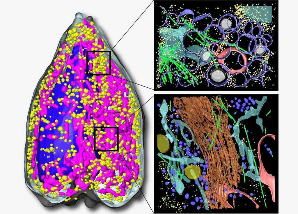
Now, scientists at the Berkeley Lab‘s Advanced Light Source have developed the world’s first soft X-ray tomography (SXT) microscope for whole-cell visualization. Using the microscope, they observed never-before-seen details about insulin secretion in pancreatic cells taken from rats.
Carolyn Larabell, Director of the National Center for X-ray Tomography (NCXT) and a Berkeley Lab, said, “Our data shows that SXT is a powerful tool to quantify subcellular rearrangements in response to drugs. This is an important first step for bridging the longstanding gap between structural biology and physiology.”
Larabell and the other authors note that SXT is uniquely suited to image whole cells without alterations from stains or added tagging molecules – as is the case for fluorescence imaging – and without chemically fixing sectioning them, which is necessary for traditional electron microscopy. Also, SXT has a much faster and easier cell preparation process.
Using their approach, scientists could visualize isolated insulin-secreting cells (called beta cells) before, during, and after stimulation from exposure to differing levels of glucose and an insulin-boosting drug. In rats and other mammals, beta cells respond to rising blood glucose levels by releasing insulin. This hormone regulates glucose metabolism throughout the body.
Larabell said, “We found that stimulating beta cells induced rapid changes in the numbers and molecular densities of insulin vesicles – the membrane ‘envelopes’ that the insulin is stored in after production. This was surprising because we expected that we should see fewer vesicles during secretion when they are emptied outside the cell. But what we observe is a rapid maturation of existing immature vesicles.”
This work was done in collaboration with researchers dedicated to whole-cell modeling, called the Pancreatic β-Cell Consortium.
Journal Reference:
- Kate L. White et al. Visualizing subcellular rearrangements in intact β cells using soft x-ray tomography. DOI: 10.1126/sciadv.abc8262