
According to two new studies, we need to reconsider the images depicting DNA is organized in the cell’s nucleus. Explaining it is essential since DNA’s spatial arrangement in the nucleus can influence the expression of genes contained inside the DNA atom, and subsequently, the proteins found in the cell.
Scientists at the Weizmann Institute of Science studied the influence of mechanical forces in the cell nucleus within the muscle. They found that muscle contraction had an immediate effect on gene expression patterns.
Prof. Talila Volk of the Molecular Genetics Department said, “We couldn’t explore this further because existing methods relied on imaging of chemically preserved cells, so they failed to capture what happens in the cell nuclei of an actual working muscle.”
This issue was addressed using a device that allowed scientists to study muscle nuclei in live fruit fly larvae. The device holds the tiny, translucent larva within a groove that permits it to contract and relax its muscles. This also keeps its movement constrained so a fluorescence microscope can scan it.
By doing so, scientists were able to obtain images of the internal, linearly-organized complexes of DNA and its proteins (known as chromatin), surrounded by the membrane of the muscle nuclei.
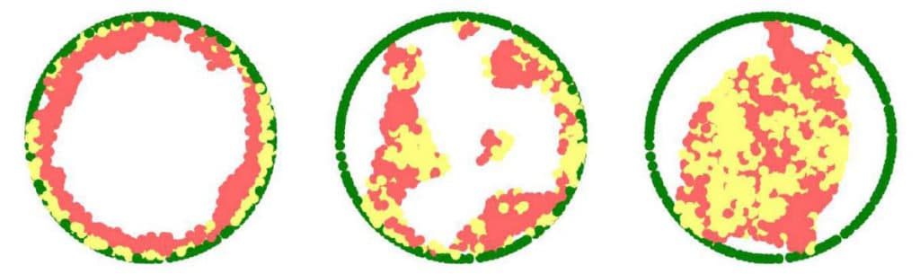
They found that- instead of filling up the entire volume of the nucleus, the ‘noodles,’ or long chromatin molecules, were organized as a relatively thin layer attached to its inner walls. Like phase separation, the chromatin separated itself from the bulk of the liquid inside of the nucleus. It found its place at its outskirts.
These findings also addressed a fundamental biological question- how is chromatin, and hence DNA, organized in the nucleus in a living organism.
Scientists also developed a theoretical model that included the physical factors governing chromatin organization in the nucleus, such as the relative forces of attraction between chromatin and its liquid environment and between chromatin and the nuclear membrane.
The model anticipated that the chromatin should go through partition from the liquid phase, contingent upon the relative amount of liquid (hydration) in the nucleus. Besides, the phase-separated chromatin could then arrange itself along within the atomic film.
Prof. Sam Safran of the Chemical and Biological Physics Department at the Weizmann Institute of Science said, “The groups also explained why in previous studies by other scientists, the chromatin appeared to fill the cell nuclei. When scientists plate cells on a glass slide to study them under a microscope, they change their volume and physically flatten them. This may perturb some of the forces governing chromatin arrangement and reduce the distance between the upper part of the nucleus to its base.”
Scientists also examined live human white blood cells. This was done to ensure that the findings were not limited to fruit fly muscle cells.
Amiad-Pavlov said, “This showed that what we’d found was likely to be a general phenomenon and that this chromatin organization had probably been conserved throughout evolution.”
The study opens up new avenues of research into DNA’s organization in the cell and the physical forces that act upon the nucleus and chromatin that can affect gene expression. One potential direction is exploring whether there’s a difference between DNA organization in health and disease. If so, this difference may be exploited in diagnosis, for example, as a new parameter for detecting cancer cells.
Journal Reference:
- Daria Amiad-Pavlov et al., Live imaging of chromatin distribution reveals novel principles of nuclear architecture and chromatin compartmentalization, Science Advances (2021). DOI: 10.1126/sciadv.abf6251
- Gaurav Bajpai et al., Mesoscale phase separation of chromatin in the nucleus, eLife (2021). DOI: 10.7554/eLife.63976