The gustatory system is the sensory system responsible for the perception of taste and flavor. In mice, taste signals are relayed by multiple brain regions, including the parabrachial nucleus (PBN) of the pons, before reaching the gustatory cortex via the gustatory thalamus.
Various studies have suggested that taste information at the periphery is encoded in a labeled-line manner, such that each taste modality has its own receptors and neuronal pathway. In contrast, the molecular identity of gustatory neurons in the CNS remains unknown.
In a new study by the National Institute for Physiological Sciences in Japan, scientists have identified the neurons responsible for relaying sweet taste signals to the gustatory thalamus and cortex in mice. While the peripheral taste system has been broadly explored, moderately little is thought about the contribution of CNS gustatory neurons in the sensation of taste.
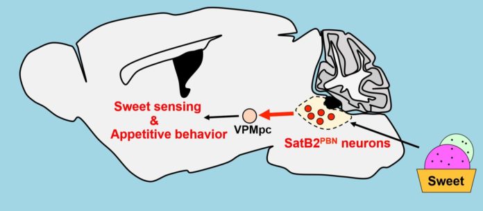
Scientists identified neurons in the brainstem that triggers sweet tastes. Particularly, scientists studied mice’s parabrachial nucleus of the pons in the brainstem, a major hub that receives sensory information about hunger, satiety and taste information. They then relay it to the cortex via the gustatory thalamus.
One clue to the molecular properties of gustatory neurons in the parabrachial nucleus may lie in the neuronal expression of SatB2; the role of neurons in the parabrachial nucleus that possesses this transcription factor has so far remained a mystery.
What scientists found that the SatB2-expressing neurons in the parabrachial nucleus of mice encode sweet tastes. They also determined that these neurons are projected to the gustatory thalamus induced appetitive lick behaviors in mice.
Ken-ichiro Nakajima at the National Institute for Physiological Sciences said, “We’ve known about the presence of taste-responsive neurons in the parabrachial nucleus for over 40 years. Only recently have we had the appropriate molecular markers and imaging methods to properly characterize these neurons—we used cell ablation, in vivo calcium imaging, and optogenetics to define the role of SatB2-expressing neurons is in the sensation of taste.”
To further evaluate the functional roles of SatB2PBN neurons in the gustatory pathway, scientists ablated these neurons by bilaterally injecting AAV expressing Cre-dependent diphtheria toxin chain A (DTA) into the PBN of SatB2-Cre mice. Doing this causes the loss of normal sweet taste sensing, which was measured by licking behavior in mice, but had little impact on the sensitivities to umami, bitter, sour, and salty tastes. In other words, it suggested that SatB2-expressing neurons have selective roles in sweet taste transduction.
Moreover, scientists cleared up the functional job of SatB2-expressing neurons. Artificial enactment by optogenetics caused dramatic changes in licking conduct; mice intensively licked tasteless water as though it were the sweet-tasting solution. These discoveries demonstrated that SatB2-expressing neurons convey sweet taste-specific signs.
Lead author Ou Fu said, “Our findings indicate that different taste qualities are processed by different types of neurons, at least in the brainstem. The next important step will be to identify a whole set of gustatory neurons, including SatB2 neurons, in the mouse parabrachial nucleus. This will allow us to understand how their assemblage forms complex flavors.”
Scientists believe that the work may help in characterizing taste processing at molecular and cellular levels.
