
Imaging brain samples involve taking little tissue tests cut into ultrathin cuts which can be recolored to uncover attributes of intrigue, for example, proteins or different markers related to infection.
In recent years, however, advances in molecular tagging techniques and the availability of laser microscopes have led to the development of modern tissue clearing techniques. Now, Imperial scientists have developed a breakthrough imaging technique which reveals the ultra-fine structure of the brain in unprecedented detail.
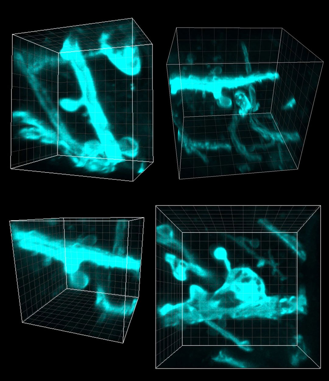
The method allows scientists to take 3D insights of the human brain, resulting in stunning images of the human brain at the microscopic level. Scientists believe that the technique could help to shed new light on the basis of neurological diseases which affect millions around the world.
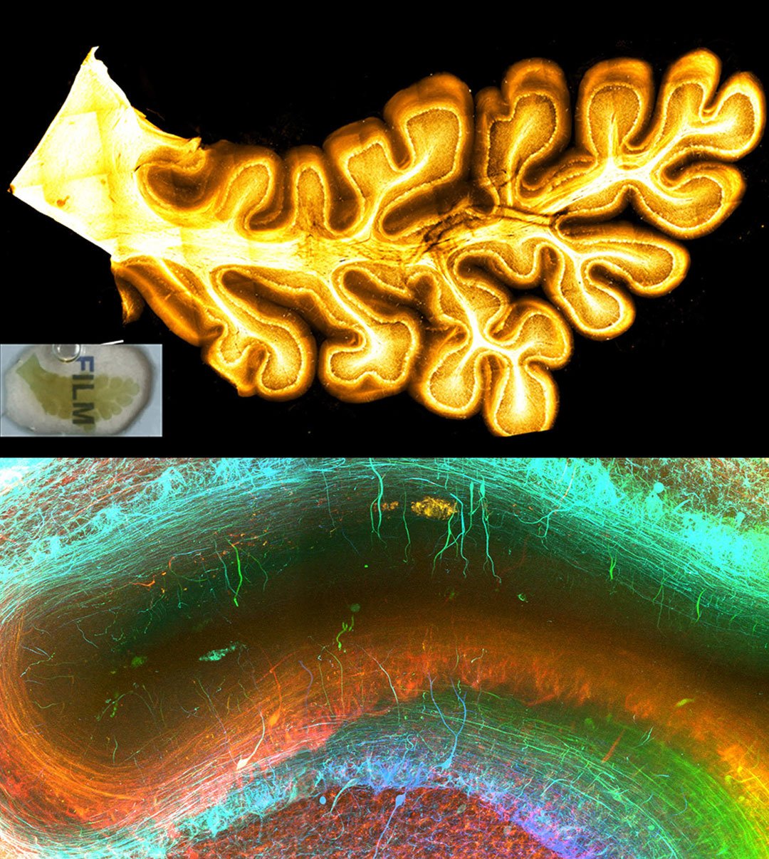
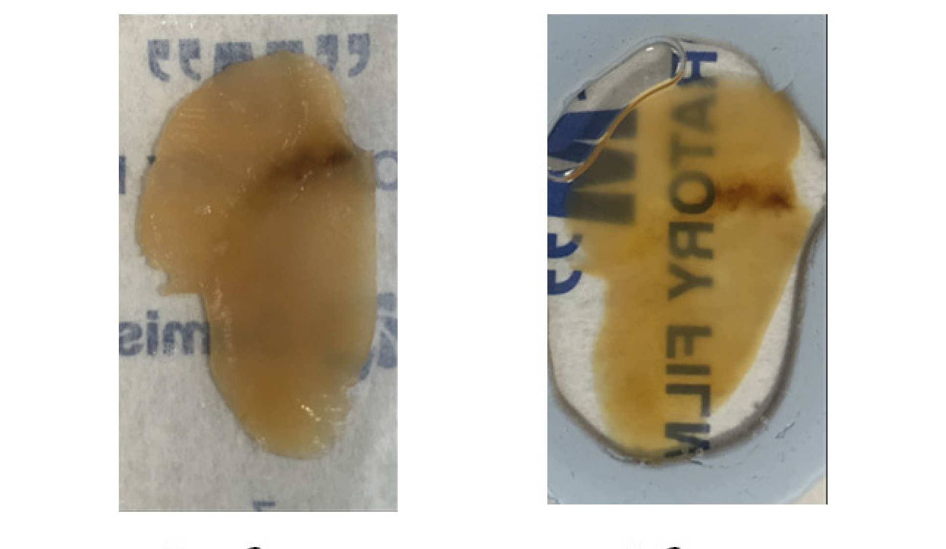
With the help of 3 medical students, scientists primarily developed a new tissue clearing solution, OPTIClear, enabling a wide range of molecular labeling methods for 3D visualization of fresh and archival human brain tissue.
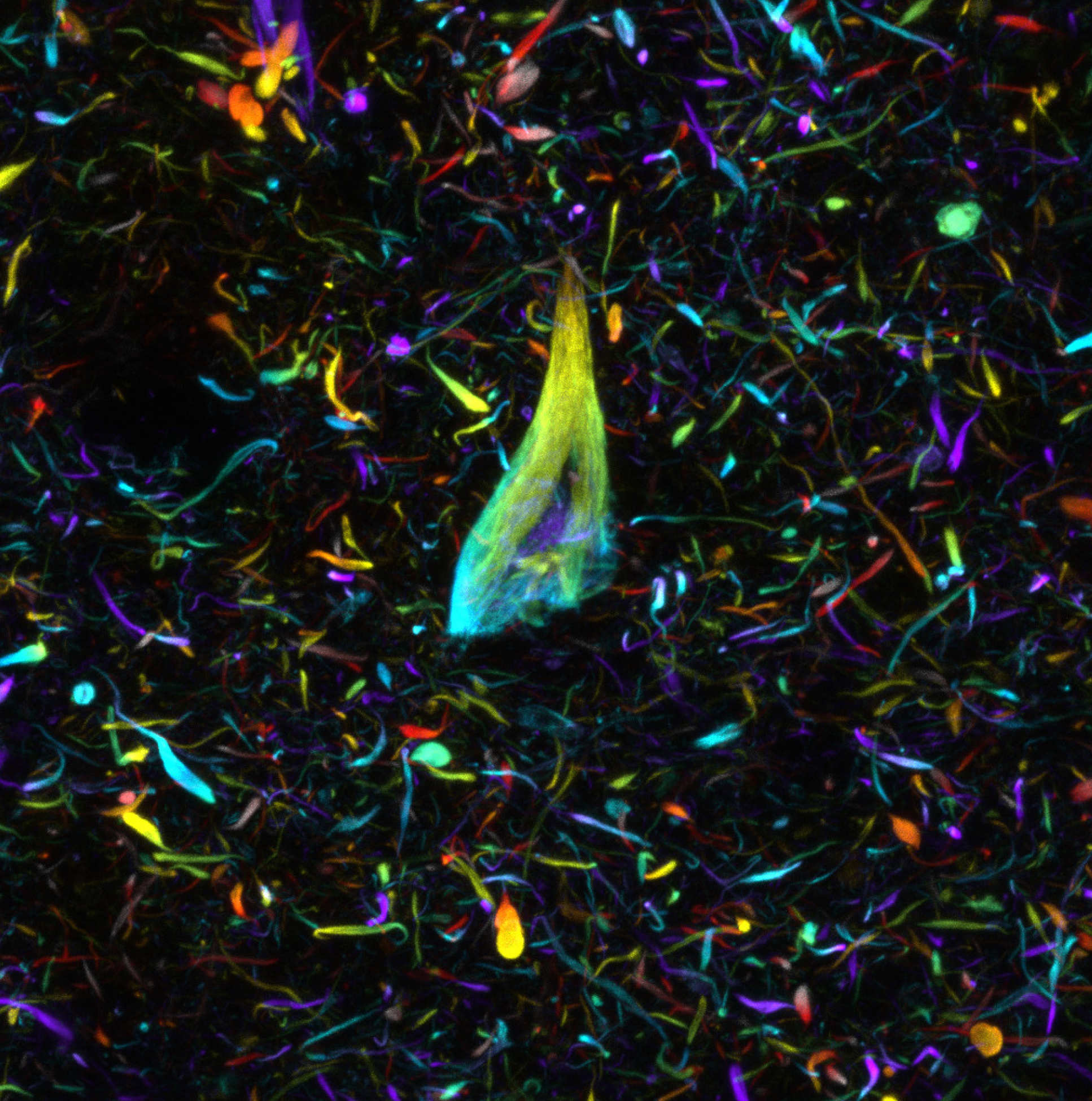
Using this new approach, they have been able to stain nerve cells, glial cells and blood vessels, as well as pathological markers such as tangles of tau protein, found in the brains of Alzheimer’s patients, in exquisite detail and determine how they relate to each other in 3D space.
Professor Steve Gentleman, Scientific Director of the Parkinson’s UK Brain Bank at Imperial College London said, “These techniques enable us to reveal the microscopic structure of the human brain in spectacular detail.”
“By using tools such as these in the lab we will be able to visualize how cells interact with each other in 3D and learn more about the pathways and connections that are damaged in the common neurodegenerative brain conditions which have such an enormous impact on people’s lives.”
As indicated by the scientists, the strategy is generally economical, is time proficient and simple to do, and is probably going to frame the reason for encouraging procedure improvement.
It is trusted that a superior comprehension of the associations and hardware of the mind at this level will help reveal the pathologies that underlie the normal degenerative ailments of the cerebrum, for example, Alzheimer’s and Parkinson’s sickness.
The work was funded by the British Neuropathological Society, Alzheimer’s Research UK and by an Innovation grant from Parkinson’s UK.
The study is published in the journal Nature Communications.