Once upon a time, growing organs in the lab were science fiction. But now, methods such as stem cell biology and tissue engineering have turned that fiction into reality with the advent of organoids.
Organoids are tiny lab-grown tissues and organs that are anatomically correct and physiologically functional.
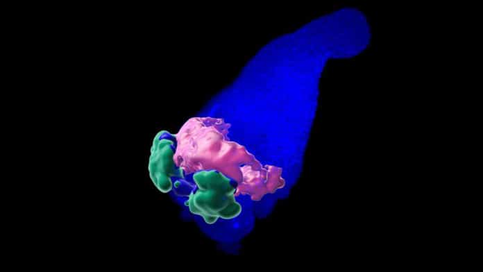
Recently, the lab of Matthias Lütolf at the School of Life Sciences at EPFL has successfully produced a mouse heart organoid in its early embryonic stages. Scientists grew organoids from mouse embryonic stem cells, which, under the right conditions, can self-organize into structures that “mimic aspects of the architecture, cellular composition, and function of tissues found in real organs.
Placed in cell-culture under specific conditions, the embryonic stem cells from a three-dimensional aggregate called a “gastruloid,” which can follow the mouse embryo’s developmental phases.
This study’s idea was that the mouse gastruloid could be utilized to mimic the beginning phases of heart development in the embryo. This is a new use of organoids, which are commonly developed to mimic adult tissues and organs.
Also, there are three features of mouse gastruloids that make them a suitable template for mimicking embryonic development: they establish a body plan like real embryos. They show similar gene expression patterns. And when it comes to the heart, which is the first organ to form and function in the embryo, the mouse gastruloid also preserves important tissue-tissue interactions necessary to grow one.
Equipped with this, the scientists exposed mouse embryonic stem cells to a “cocktail” of three factors known to promote heart growth. Following 168 hours, the subsequent gastruloids gave early heart development indications: they expressed several genes that regulate cardiovascular development in the embryo. They even generated what resembled a vascular network.
Importantly, scientists found that the gastruloids developed what they call an “anterior cardiac crescent-like domain.” This structure produced a beating heart tissue, similar to the embryonic heart. As the muscle cells of the embryonic heart, the beating compartment was also sensitive to calcium ions.
Giuliana Rossi, a post-doctoral researcher from Lütolf’s laboratory, said, “Opening up an entirely new dimension to organoids, the breakthrough work shows they can also be used to mimic embryonic stages of development. One of the advantages of embryonic organoids is that, through the co-development of multiple tissues, they preserve crucial interactions that are necessary for embryonic organogenesis.”
“The emerging cardiac cells are thus exposed to a context similar to the one that they encounter in the embryo.”
The study was conducted in collaboration with Viventis Microscopy, EPFL Bioimaging and Optics Platform, Institut de Biologie du Développement de Marseille, Johns Hopkins University School of Medicine, EPFL Institute of Chemical Sciences and Engineering.
Journal Reference:
- Giuliana Rossi, Nicolas Broguiere, Matthew Miyamoto, Andrea Boni, Romain Guiet, Mehmet Girgin, Robert G. Kelly, Chulan Kwon, Matthias P. Lutolf. Capturing Cardiogenesis in Gastruloids. Cell Stem Cell, 10 November 2020. DOI: 10.1016/j.stem.2020.10.013
