
In a new study, scientists from the University of Missouri have mapped how materials in cells travel. They determined the mechanisms underlying protein translocation remains a major unsolved problem.
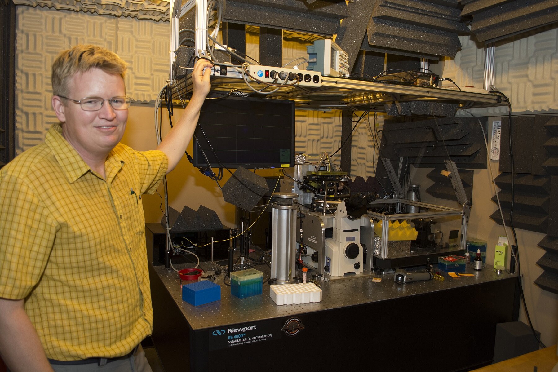
Gavin King, an associate professor of physics in the MU College of Arts and Science and joint associate professor of biochemistry said, “Proteins are like vehicles that can carry information and materials in cells. We focused on the cells of E.coli, which have two membranes. We observed that when proteins exit the cell’s inner membrane, the cell adjusts the way it transfers the protein through the channel depending on the type of protein being transported.”
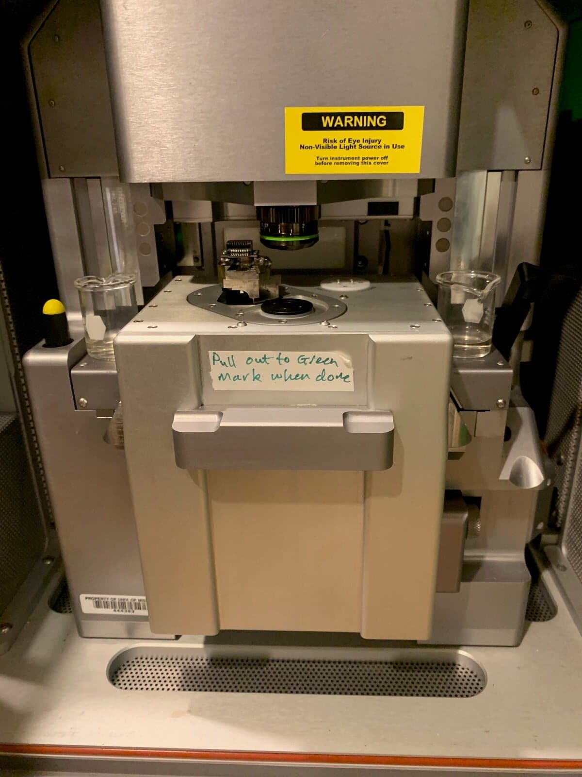
Scientists primarily verified the transportation function of E.coli cells outside their natural environment. They then sent the samples to study the movement of the E.coli proteins using an atomic force microscope. Using the microscope, scientists were able to detect the movement of proteins in a fluid environment that closely resembles their natural environment.
King said, “Before our study, there were two ways of describing how proteins moved across the cell membrane—either a piston-live movement similar to a car’s engine, or a more perpetual motion known as the Brownian ratchet. Neither model accounted for the differences based on the type of protein being transported. Just as different types of vehicles move differently so do different types of proteins. The maps we made to depict the proteins’ movement suggest nature might be more complex than previously thought.”
The study offers fundamental knowledge of how a cell sends and receives material and information. For example, how drugs pass through membranes to affect a cell.
While other cells besides E.coli may not have E.coli’s exact protein transportation system, King said a similar system exists in all cells.
The study is published in the journal of Science Advances.