
Plasmon-based devices are powerful for use in highly sensitive evanescent-field detection and analysis, but they exhibit the problem of limited frequency tunability for fixed structures. This feature causes problems in the multi-frequency investigations required for materials characterization, bio-related research, etc.
In a new study by the Tokyo Tech, scientists have proposed a new device that enables a continuous frequency-tuneable evanescent-field concentration in the terahertz (THz) region with simple operation.
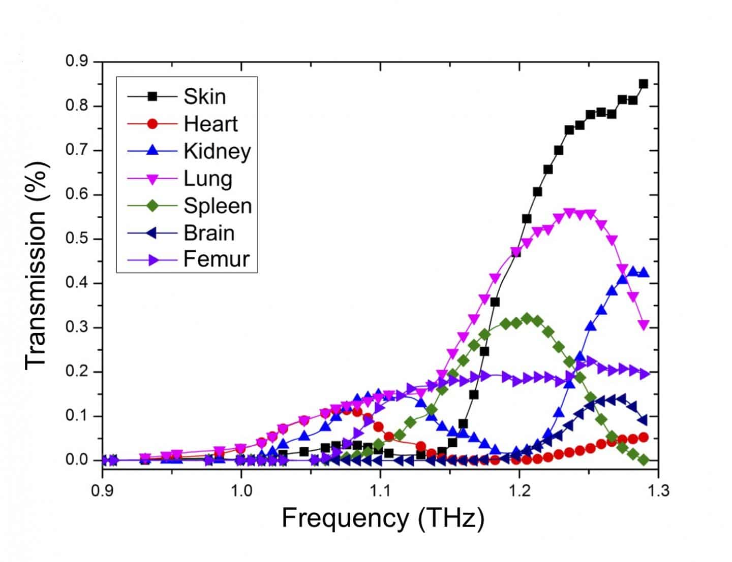
Scientists have developed an easy-to-use, tunable biosensor tailored for the terahertz range. During the experiment, this frequency-tunable plasmonic-based THz device was able to obtain the images of mouse organs. The images verified that the sensor is capable of distinguishing between different tissues.
CREDIT: Scientific Reports
One of the key features of the new device is its spiral bull’s eye (SBE) design. Due to its smoothly varied grooves, the groove period continuously changes with the diameter direction, resulting in continuously frequency-tunable characteristics.
Moreover, the device incorporates a so-called Siemens-star aperture that enables a user-friendly way of selecting the desired frequency by simply changing the rotation of the spiral plasmonic structure.
Yukio Kawano at Tokyo Tech’s Laboratory for Future Interdisciplinary Research of Science and Technology said, “The device also increases the electric field intensity at the subwavelength aperture, thus significantly amplifying the transmission.”
CREDIT
Scientific Reports
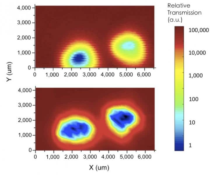
To probe further, they also conducted THz mapping of mouse tails. By comparing images obtained with and without the SBE design, the study showed that the former led to a markedly improved ability to distinguish between different tissues such as hair, skin, and bone.
Kawano said, “Further investigations are planned to test the new device using various mouse organ tissues. The findings open up a new direction for plasmonic-based THz imaging of biological samples, which may eventually lead to the development of improved, non-invasive diagnostic imaging tools.”
The study is published in Scientific Reports.