The zebrafish (Danio rerio) is an important vertebrate model organism. One of the most readily observable phenotypes in the zebrafish is its distinctive pigmentation patterning, which, in wild-type adults, appears as alternating dark and light longitudinal stripes.
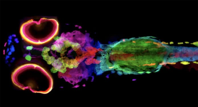
Scientists at Berkeley Lab have developed a new technique to image every pigment cell of a whole zebrafish in 3D.
Melanin is a natural skin pigment. But studying melanin directly with a conventional microscope is challenging. Hence, scientists turned to X-ray imaging, which can pass through optically opaque matter like melanin.
Scientists performed imaging on two sets of zebrafish samples – one with the normal pigmentation associated with the zebrafish’s characteristic black stripes and another from a mutant zebrafish line with lighter stripes called golden.
They then visualized the melanin by using a newly developed staining technique. The technique binds to the pigment and blocks X-rays.
Using the ALS and an X-ray detector system, scientists visualized every melanocyte – a cell containing melanin – of whole wild-type and golden zebrafish larvae about 1.5 mm long. They then used the micro-computed tomography (micro-CT) technique to map the cells in 3D.
The technique combines the new detector system with high X-ray flux from the ALS. Doing so, they achieved a resolution that is 2,000-fold higher than conventional CT.
Scientists noted, “The silver-staining technique could be used to learn more about the 3D architecture of melanoma tumors and potentially guide treatment decisions.”
Journal Reference:
- Spencer R Katz et al. Whole-organism 3D quantitative characterization of zebrafish melanin by silver deposition micro-CT. DOI: 10.7554/eLife.68920
