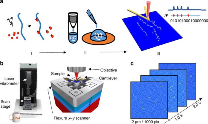
Bristol scientists have supercharged their atomic force microscope (AFM) in order to map DNA mutations in the human body. They believe that their technology could transform the way of diagnosing disease-causing genetic mutations.
Scientists used off-the-shelf DVD components to enable it to enable it to physically map the lengths of individual DNA molecules to a resolution of tens of base pairs at rates of hundreds per second.
This phenomenal speed increment empowers this DNA barcoding strategy to be utilized for genuine diagnostics out of the blue.
This nanomapping microscope can map almost hundreds of chemically barcoded DNA molecules every second.
Scientists used CRISPR-Cas9 to mark the particles with the goal that they can be mapped practically as precisely as DNA sequencing, while additionally handling huge segments of the genome at a considerably quicker rate.
The AFM technology was introduced in 1989 by IBM scientists. At that time, they used the technique to rearrange molecules at the atomic level to spell out ‘IBM’.
The microscope measures single DNA particles with sub-nuclear determination while making pictures up to a million base combines in an estimate. What’s more, it does it utilizing a small amount of the measure of example required for DNA sequencing, drastically lessening the estimation time.
Dr Oliver Payton from the University of Bristol said, “Using the laser focusing mechanism found in every DVD player we have built a microscope that has the resolution and speed to measure every molecule on the sample surface in 3D.”
“Although other types of microscope have the resolution to see these DNA molecules they are thousands of times slower and it would take years to make a confident diagnosis.”
“Not only is our microscope perfect for these medical applications, but because of the readily available DVD player components it can be mass-produced.”
To test the system’s efficiency, specialists mapped hereditary translocations display in lymph hub biopsies of lymphoma patients.
Translocations happen when one area of the DNA gets reordered to the wrong place in the genome. They are particularly predominant in blood growths, for example, lymphoma yet happens in different diseases too.
Professor Reed said, “Because the CRISPR enzyme is a protein that’s physically bigger than the DNA molecule, it’s perfect for this barcoding application.”
“We were amazed to discover this method is nearly 90 percent efficient at binding to the DNA molecules. And because it’s easy to see the CRISPR proteins, you can spot genetic mutations among the patterns in DNA.”
Now scientists are using their nanomapping microscope to solve a diverse range of nanoscale challenges across fields such as 2D materials, corrosion and life sciences.
