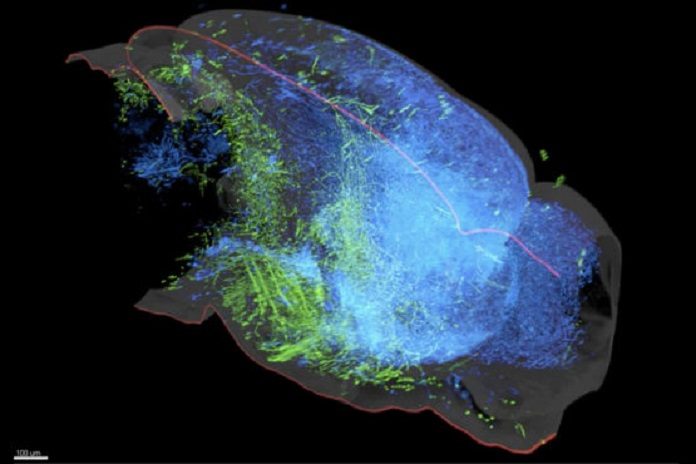
Earlier before six years, using neuroanatomical methods that they invented, scientists found that the brain’s serotonin system in brain promotes pleasure. Now, after six years, Stanford scientists found that the serotonin system is actually composed of at least two, and likely more, parallel subsystems that work in concert to affect the brain in different, and sometimes opposing, ways.
Scientists have created a detailed map of the brain’s serotonin system. Researchers were finding particular elements of serotonin in the brain and ascribing them to a monolithic serotonin system, which at any rate mostly represents the controversy about what serotonin really does.
Scientists expect that this examination enables them to see diverse parts of the elephant in the meantime. In addition, it could have implications for the treatment of depression and anxiety, which involves prescribing drugs such as Prozac that target the serotonin system – so-called SSRIs (selective serotonin reuptake inhibitors).
During this study, scientists concentrated on a region of the brainstem known as the dorsal raphe, which contains the biggest single focus in the mammalian brain of neurons that all transmit signals by discharging serotonin.
The nerve fibers, or axons, of these dorsal raphe neurons, convey a sprawling system of connections with numerous many critical forebrain regions that complete a large group of capacities, including considering, memory, and the control of states of mind and substantial capacities. Scientists injected viruses to infect serotonin axons and traced the connections back to their origin neurons in the dorsal raphe.
Doing this, they created a visual map of projections between the dense concentration of serotonin-releasing neurons in the brainstem to the various regions of the forebrain that they influence. The guide uncovered two particular groups of serotonin-releasing neurons in the dorsal raphe, which associated with cortical and subcortical areas in the brain.
In a series of behavioral tests, the scientists also showed that serotonin neurons from the two groups can respond differently to stimuli. For example, neurons in both groups fired in response to mice receiving rewards like sips of sugar water but they showed opposite responses to punishments like mild foot shocks.
Liqun Luo, who is the Ann and Bill Swindells Professor in the School of Humanities and Sciences at Stanford University said, “We now understand why some scientists thought serotonin neurons are activated by punishment, while others thought it was inhibited by punishment. Both are correct – it just depends on which subtype you’re looking at.”
“We also found that the serotonin neurons themselves were more complex than originally thought. Rather than just transmitting messages with serotonin, the cortical-projecting neurons also released a chemical messenger called glutamate – making them one of the few known examples of neurons in the brain that release two different chemicals.”
Study first author Jing Ren said, “It raises the question of whether we should even be calling these serotonin neurons because neurons are named after the neurotransmitters they release.”
Taken together, these findings indicate that the brain’s serotonin system is not made up of a homogenous population of neurons but rather many subpopulations acting in concert. Luo’s team has identified two groups, but there could be many others.
In fact, Robert Malenka, a professor and associate chair of psychiatry and behavioral sciences at Stanford’s School of Medicine, and his team recently discovered a group of serotonin neurons in the dorsal raphe that project to the nucleus accumbens, the part of the brain that promotes social behaviors.
