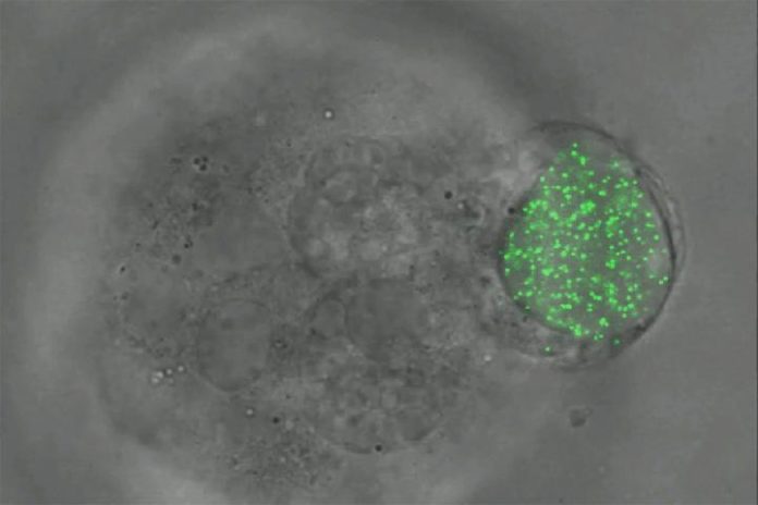
Scientists at the University of Illinois have developed a new technique that uses tiny elastic balls filled with fluorescent nanoparticles aims to expand the understanding of the mechanical forces that exist between cells. Using the technique, scientists show the evaluation of 3-D forces called traction inside cells living in Petri dishes and in addition live examples.
The magnitudes of these mechanical forces are far from trivial at the cellular level. The study suggests that the traction plays a fundamental role in cell physiology.
As cells develop and recreate, they apply force to each other while going after space. The group found that in the event that they infuse their modest flexible spheres into beginning time incipient organisms of zebrafish and settlements of melanoma cells of mice in Petri dishes, they too encounter the powers.
Ning Wang, research led said, “If we place a single cell in a medium within a petri dish it will not survive for long, even if we provide all of the nutrients needed. The cells fail to form any sort of tissue because there is no support or scaffolding on which to build.”
In order to quantify the amount of force imposed on the cells, scientists placed fluorescent nanoparticles inside of the spheres. At the point when the cells crush the spheres, the nanoparticles all move the same amount per area of force. The specialists would then be able to quantify the movements of the glowing particles utilizing fluorescent microscopy to figure the measure of power applied to the circles and cells. Utilizing this method, the group has denoted the principal fruitful estimation of each of the three sorts of power – pressure, strain, and shear – in every one of the three measurements.
Wang reported, “We thought that cancer cells would generate more pressure at this early growth stage while the mass of the tumor increases, as we observed in zebrafish embryos, but they do not. We suspect that the cancer cells begin to spread out or metastasize right after this stage.”
“Although the underlying mechanism for metastasis is unclear, we have hypothesized that tumor-repopulating cells spread very rapidly in these secondary soft tissues. Having the ability to measure changes in tractions at the intercellular level may serve as an early cancer-detection tool.”
This microgel sphere technology may also help unravel the mechanisms behind a metastasis-halting synthetic drug. Scientists are continuing to apply this technology to stem and embryonic cell research.
Scientists reported their findings in the journal Nature Communications.
