
Norovirus is a very contagious virus that causes vomiting and diarrhea.
To understand the structure of the norovirus, scientists from the University of Leeds created better visualization of a strain of the norovirus from 13,000 separate images taken by an electron microscope.
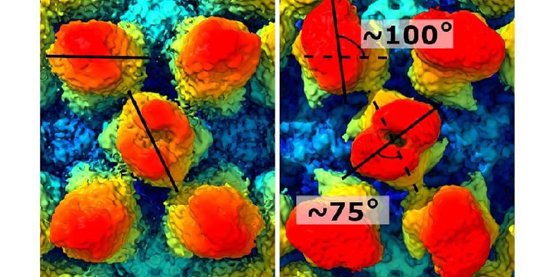
The visualization reveals in rich detail the structure of the virus-like protrusions on the outside of the virus capsid, the protein shell that holds the genome.
Joseph Snowden, a Ph.D. researcher from the Astbury Centre for Structural Molecular Biology, said, “it revealed that the protein casing did not keep a fixed shape, as the protrusions on the surface would extend, retract and rotate.”
For the study, scientists used mouse norovirus, which is similar to human norovirus.
Using a method called focussed classification, scientists identified ultra-fine changes to the structure of these protein shells.
Scientists found that the protrusions move independently or in a coordinated fashion. According to scientists, this may enable the virus to prime itself ready for infection depending on where it is, for example, if it is in the digestive tract of a host organism.
Mr. Snowden said: “The constant changing or morphing of the virus shell may enable it to confuse the body’s defense systems.
“Immune systems work based on molecular shape. The body’s defenses will produce proteins that bind to pathogens to prevent them from infecting host cells. But if the shape of the virus is constantly changing, the body’s defense systems may be unable to act efficiently.”
Dr. Morgan Herod, from the School of Molecular and Cellular Biology at Leeds and one of the senior authors on this study, hopes that a greater understanding of the structure of the norovirus may help with vaccine development, which so far has been unsuccessful.
Dr. Herod said, “The University of Leeds has a strong track record in the use of virus-like particles (VLPs) to create vaccine candidates, for example, against polio. VLPs are harmless proteins modeled on the structure of a virus’s casing, and they fool the immune system into thinking the body is under attack, prompting an immune response.”
“Our research shows that the capsid or protein shell of norovirus is dynamic – and perhaps we have to look at VLPs that are better able to mimic this aspect of norovirus structure.”
“That will make the vaccine development task a little harder.”
Journal Reference:
- Dynamics in the murine norovirus capsid revealed by high-resolution cryo-EM. DOI: 10.1371/journal.pbio.3000649