
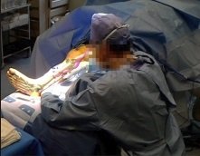
For the first time, surgeons have used Microsoft HoloLens headsets while operating on patients to undergo reconstructive lower limb surgery. Researchers have revealed this in a series of procedures are carried out by a team at Imperial College London at St Mary’s Hospital.
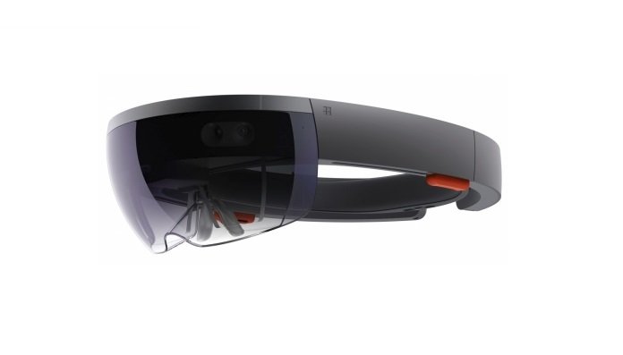
These HoloLens actually are self-contained computer headset that immerses the wearer in ‘mixed reality’, enabling them to interact with ‘holograms’ (computer-generated objects made visible through the visor).
© Imperial College London
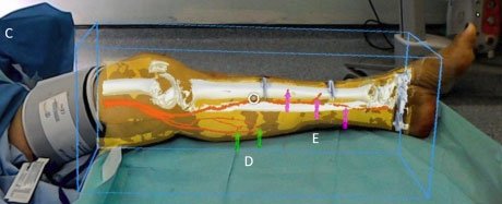
Researchers used this technology to overlay images of CT scans, including the position of bones and key blood vessels, onto each patient’s leg, in effect enabling the surgeon to ‘see through’ the limb during surgery.
This novel approach can help surgeons locate and reconnect key blood vessels during reconstructive surgery, which could improve outcomes for patients.
Dr. Philip Pratt a Research Fellow in the Department of Surgery, said, “We are one of the first groups in the world to use the HoloLens successfully in the operating theatre.”
“Through this initial series of patient cases, we have shown that the technology is practical and that it can provide a benefit to the surgical team. With the HoloLens, you look at the leg and essentially see inside of it. You see the bones, the course of the blood vessels, and can identify exactly where the targets are located.”
© Imperial College London
In many cases like a car accident or severe trauma, patients may have tissue damage or open wounds that require reconstructive surgery using fasciocutaneous flaps. These flaps of tissue, which are taken from elsewhere on the body and include the skin and blood vessels, are used to cover the wound and enable it to close and heal properly.
A crucial step in the process is connecting the blood vessels of the ‘new’ tissue with those at the site of the wound, so oxygenated blood can reach the new tissue and keep it alive.
The standard approach for this element of reconstructive surgery has been the use of a handheld scanner which uses ultrasound to identify blood vessels under the skin by detecting the movement of blood pulsing through them, enabling the surgeon to approximate where the vessels are and their course through the tissue.
Dr. Pratt explained, “Augmented reality offers a new way to find these blood vessels under the skin accurately and quickly by overlaying scan images onto the patient during the operation.”
In the trial of this technology, five patients requiring reconstructive surgery on their legs underwent CT scans to map the structure of the limb, including the position of bones and the location and course of blood vessels.
© Imperial College London
Dr. Dimitri Amiras, a consultant radiologist at Imperial College Healthcare NHS Trust (ICHNT) segmented the images from the scans into bone, muscle, fatty tissue and blood vessels whereupon loaded into the intermediary software to create 3D models of the leg.
These models were then fed into specially designed software that renders the images for the HoloLens headset, which in turn overlays the model onto what the surgeon can see in the operating theatre.
After which clinical staff is able to manipulate these augmented realities (AR) images through hand gestures to make any fine adjustments and correctly line up the model with surgical landmarks on the patient’s limbs, such as the knee joint or ankle bone.
Dr. Amiras said, “St Mary’s Hospital is a major trauma center, giving us the opportunity to try and improve the pre-operative planning for reconstructive flaps.
“Over time, the scanning protocol has been optimized to give excellent images of the anatomy, however, at first we had to rely on rough measurements of anatomical landmarks taken from 3D CT reconstructions to guide surgery.”
“Now, using the HoloLens, we can identify where the blood vessels are in 3D space and use virtual 3D arrows to guide the surgeon.
“Currently, data preparation is a time-consuming process, but in the future, much of this could be automated, with the consultant radiologist checking the accuracy of the model against the original scan.
“I think this is a great example of what can be achieved in an Academic Health Science Centre.”
In real life, cases ranged from a 41-year-old man who had sustained leg injuries during a car accident, to an 85-year-old woman with a compound fractured ankle.
The surgical teams reported the HoloLens to be a powerful tool in the theatre, with the approach being more reliable and less time-consuming than the ultrasound method of locating blood vessels.
Mr. Jon Simmons, a plastic and reconstructive surgeon at ICHNT, said, “The application of AR technology in the operating theatre has some really exciting possibilities.”
“While the technology can’t replace the skill and experience of the clinical team, it could potentially help to reduce the time a patient spends under anesthetic and reduce the margin for error. We hope that it will allow us to provide more tailored surgical solutions for individual patients.”
The team of researchers highlights a few limitations with the technology, which could include errors during the modeling stages as well as the potential for the overlaid model to be misaligned.
In addition, the case studies so far have been based on the leg, which has a number of clearly visible surgical ‘landmarks’, such as the ankle or knee.
Areas without these rigid landmarks, such as the abdomen, may be more complicated with a greater potential for movement of blood vessels.
However, the researchers are confident that, once refined, the approach could be applied to other areas of reconstructive surgery requiring tissue flaps, such as breast reconstruction following mastectomy.
The next steps include trialing the technology in a larger set of patients, with procedures carried out by teams at multiple centers.
Dr. Pratt added, “In future, we hope to automate the process further. We can use software to improve the alignment and will attach markers to the patient when they have the scan, with the same markers present during the operation to use as additional points of reference.
He added, “There are a number of areas we would like to explore, and further improvements are needed, but the small case series have shown that for reconstructive surgery, this seems to be a valuable tool in the operating theatre.”
‘Through the HoloLens looking glass: augmented reality for extremity reconstruction surgery using 3D vascular models with perforating vessels’ by Philip Pratt et al. is published in the journal European Radiology Experimental.