
Quantitative examination of plant and animal morphogenesis requires the precise segmentation of individual cells in volumetric pictures of developing organs. In the most recent years, deep learning has given vigorous automated algorithms that approach human performance, with bio-image analysis applications now beginning to develop.
In a new study, an EMBL research group collaborated with the DFG-funded FOR2581 consortium and developed a new tool based on machine learning that could tackle plant cell jigsaw puzzle. The tool called PlantSeg offers the most accurate and versatile analysis of plant tissue development to date.
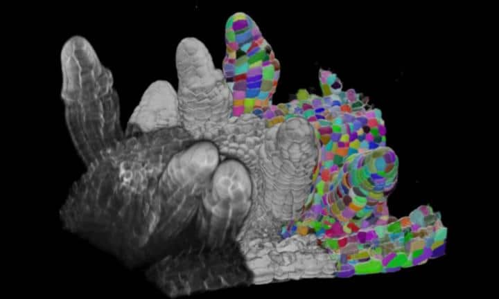
The PlantSeg take a 3D perspective of cells and separate them all is surprisingly hard to do, considering how easy it is for humans.
Anna Kreshuk, a computer scientist and expert in machine learning, said, “Computers aren’t as good as humans when it comes to most vision-related tasks, as a rule. With all the recent development in deep learning and artificial intelligence at large, we are closer to solving this now, but it’s still not solved – not for all conditions. This paper is the presentation of our current approach, which took some years to build.”
“In plants, you have cells that look extremely regular that in a cross-section looks like rectangles or cylinders. But you also have cells with so-called ‘high lobeness’ that have protrusions, making them look more like puzzle pieces. These are more difficult to segment because of their irregularity.”
Scientists trained PlantSeg on 3D confocal images of fixed Arabidopsis thaliana ovules and 3D+t light-sheet microscope images of developing lateral roots, two standard imaging modalities in plant morphogenesis studies. They explored a range of network architectures and graph partitioning algorithms and selected the ones who performed best with regard to the extensive, manually annotated ground truth.
Benchmarking PlantSeg on different datasets covering a range of samples and image resolutions, scientists found that the tool delivers excellent results on unseen data.
Kreshuk said, “We have giant puzzle boards with thousands of cells, and then we’re essentially coloring each one of these puzzle pieces with a different color.”
“This kind of algorithm allows for all kinds of shape-related analysis, for example, analysis of shape changes through development or under a change in environmental conditions, or between species. The paper gives some examples, such as characterizing developmental changes in ovules, studying the first asymmetric cell division which initiates the formation of the lateral root, and comparing and contrasting the shape of leaf cells between two different plant species.”
“While this tool currently targets plants specifically, but it could be tweaked to be used for other living organisms as well.”
Journal Reference:
- Adrian Wolny et al. Accurate and versatile 3D segmentation of plant tissues at cellular resolution. DOI: 10.7554/eLife.57613