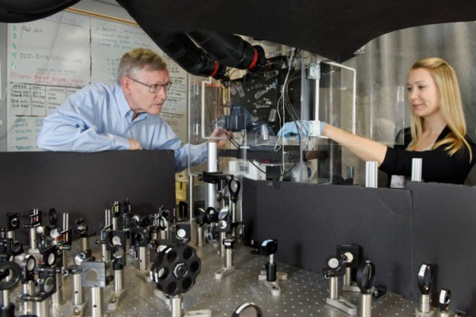
Standford scientists have recently won the Nobel Prize in chemistry for their innovation of imaging shapes inside cells at very high resolution, called super-resolution microscopy. Now, scientists have developed a new microscope that produces 3-D nanoscale images of mammalian cells in their entirety.
W. E. Moerner, Professor of Chemistry at Stanford University said, “A cell has a whole city of proteins, enzymes and structures working all the time. We have some idea of what’s in a cell – many of us are familiar with drawings of mitochondria or of the endoplasmic reticulum – but it’s an average idea. When we look at individual cells, we recognize that they all aren’t exactly like the pictures we have in textbooks.”
Scientists dubbed this microscope as TILT3D that combines two new imaging techniques with super-resolution microscopy to capture very clear 3-D images of structures and individual molecules within a cell.
In most of the light microscopes, the cell sample is lit from below. This is an issue on the off chance that you need to explore the subtle elements of a cell since it prompts outwardly dim pictures where just a few sections are in a center – like a photograph assumed control over a long separation.
Standard light sheet brightening gets around this issue by sparkling just a cut of light in from the side to get a hotcake like enlightenment of the example. Indeed, even with this preferred standpoint, on the off chance that you attempt to get a light sheet to sparkle on the exceptionally base of a cell, it ricochets off the side of the chamber containing the example, which twists the picture. By tilting the light sheet, the Moerner lab abstains from hitting the corner.
Notwithstanding clearing the visual mess by tilting the light sheet, the new magnifying instrument incorporates an optical strategy for imaging in 3-D. To accomplish this, the scientists label atoms in the cell test with chemicals that fluoresce when lit and utilize concoction added substances to influence them to flicker splendidly. At that point, through what Moerner calls “optical enchantment,” the gathering alters the magnifying instrument to change over every fluorescent squint into two spots of light at various edges. With these two spots, the specialists can get the situation of every atom in three measurements, which educates the last 3-D picture.
Stacking their pancaked 3-D pictures over each other, the specialists can make a through and through a recreation of a cell. Tilted light sheet imaging likewise makes it conceivable to track the 3-D development of particles after some time with an accuracy of several nanometers, which could catch atoms holding, moving by engines or voyaging arbitrarily through structures of the cell.
Joining TILT3D’s unmistakable picture and 3-D capacities with existing super-determination strategies, the magnifying instrument can make exact pictures at super-determination – as little as many nanometers or around 4,000 times littler than a human hair is thick. This opens up new chances to deliver definite 3-D pictures of mammalian cell structures, even of ones that were beforehand excessively thick, making it impossible to picture unmistakably.
Scientists tested their microscope on known cell structures. They are as of now strolling different labs through the way toward copying this magnifying instrument. The plan can be a measured expansion of existing light magnifying instruments. Later on, they trust that their 3-D tilted light sheet enlightenment imaging will be utilized for any number of undertakings.
Gustavsson, who is partly supported by a postdoctoral fellowship from the Karolinska Institutet in Sweden said, “TILT3D is simpler than other microscopes that have been designed for imaging of these challenging samples, and it can be used for imaging both of static structures and of moving molecules. We designed it to be versatile, not bound to a specific question.”
The analysts will keep on working on TILT3D, especially on joining static and dynamic data from a few distinct proteins. Close by their numerous different developments and concentrates in cell imaging, they trust this innovation can empower them and others to take in more about the structures and procedures of cells, one particle at any given moment.
The microscope was described recently in a paper published in Nature Communications.
