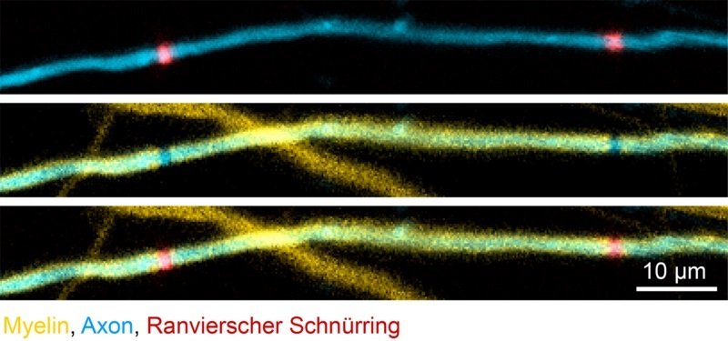
Nerve fibers are encompassed by a myelin sheath. Researchers at the Technical University of Munich (TUM) have now made the first-ever “live” observations of how this defensive layer is shaped. The group found that the trademark examples of the myelin layer are resolved at a beginning period. Be that as it may, these examples can be balanced as required in a procedure clearly controlled by the nerve cells themselves.
The myelin sheath encompassing the axons, or nerve filaments, can be contrasted with the protective covering an electric wire. Without it, the quick proliferation of electric signs would not be conceivable. Harm to this protecting layer, for instance from diseases, for example, different sclerosis, may bring about genuine impedances.
Myelin does not form a continuous coating along the axon. Instead, it is divided into segments. These can vary in length and are separated by gaps known as nodes of Ranvier. For the complex network of the central nervous system to function properly, the speed of the connections is not the only consideration.
The vital factor is the tweaking: the signs need to achieve the correct place at precisely the opportune time. The transmission speed of data through an axon is incompletely controlled by the number and length of the portions.
The collections of people and creatures have the capacity – at any rate to some degree – to repair harmed myelin sheaths. Dr. Tim Czopka, a neuroscientist at TUM, has prevailed with regards to making the first-since forever “live” perceptions of this procedure. Czopka and his group utilized recently created markers to envision the development myelin fragments encompassing axons in the spinal string of zebrafish.
They finished up: Characteristic examples made up of myelin portions with various lengths along an axon are characterized inside a couple of days after myelin development starts. Despite the fact that the fragments keep developing from that time forward – as the body of the zebrafish develops – the example stays saved.
Following this perception, the scientists wrecked chose fragments. “What occurred next shocked us,” says Tim Czopka. “After the decimation of the sections, the myelin sheaths started to powerfully rebuild. At last, the harm was repaired and by and large the first example was reestablished.”
The recovery took after a settled succession: First, the neighboring sections extended to close the hole. Another fragment at that point framed amongst them, and they contracted to their unique size.
Tim Czopka said, “This raises an important question: Who controls the emergence and the restoration of the segment pattern? Our observations suggest that it is not the oligodendrocytes – the cells that form myelin – that decide where it is formed, but rather the axons. You could say that they know best which pattern is needed for the signals to be transmitted at optimal speed.”
“The team is now studying how the segment patterns are affected by the targeted stimulation of nerve cell activity and by the neurotransmitters released as a result. If we can understand the role of the axons in myelin generation and remodeling, it may yield new approaches to controlling it. That would be relevant for the treatment of illnesses such as multiple sclerosis.”
