
Every year more than four million people die of severe respiratory ailments worldwide. Although advanced medical imaging technologies give detailed diagnostic information, a low-dose, fast, and inexpensive option for early detection of respiratory diseases and follow-ups is still lacking.
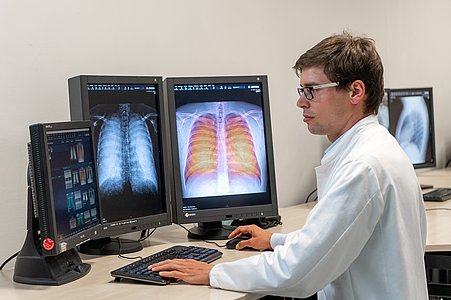
Scientists at the Technical University of Munich have developed a procedure to fill this gap: dark-field chest X-rays. Until now, this method has not been studied in living humans. For the first time, scientists at TUM have successfully used this new X-ray method for respiratory diagnostics with patients.
This new x-ray technology visualizes early changes in the alveolar structure caused by the lung disease COPD. It uses only one-fiftieth of the radiation dose typically applied in X-ray computed tomography.
Unlike conventional X-ray imaging, which is based on attenuation of X-rays on their way through the tissue, This new x-ray technology uses the wave nature of X-ray light. It mainly uses the physical phenomenon of scattering like the long-known principle of dark-field microscopy with visible light.
This phenomenon allows visualizing the structure of objects that are, for the most part, transparent. These structures appear in the microscope as bright images on a dark background, which has given the method its name.
Franz Pfeiffer, who led the study, said, “The X-ray dark-field signal is particularly strong for interfaces between air and tissue. This makes it possible for a dark-field X-ray image of the lung to clearly distinguish between intact alveoli, i.e., those filled with air, and regions in which less intact alveoli exist.”
“We expect the radiation exposure to be reduced by a factor of fifty. Furthermore, the first clinical results have confirmed that the dark-field X-rays provide additional image information on the underlying microstructure of the lung.”
Dr. Alexander Fingerle, senior physician at TUM’s university hospital Klinikum Rechts der Isar’s Department of Diagnostic and Interventional Radiology, said, “Given the close connection between the alveolar structure and the functional condition of the lung, this ability is of great significance for pulmonary medicine. In the future, dark-field X-rays could help improve early detection of COPD and other respiratory ailments.”
The clinical trials offer hope for further clinical studies and the development of marketable devices that use the dark-field method.
Pfeiffer said, “Dark-field chest X-rays are currently giving us a chance to improve the early detection of lung diseases significantly and at the same time to implement it on a wider basis than before.”
Journal Reference:
- Konstantin Willer, Alexander Fingerle et al. X-ray dark-field chest imaging for detecting and quantifying emphysema in patients with chronic obstructive pulmonary disease: a diagnostic accuracy study. Lancet Digital Health, Volume 3, ISSUE 11, e733-e744, November 01, 2021 – DOI: 10.1016/S2589-7500(21)00146-1