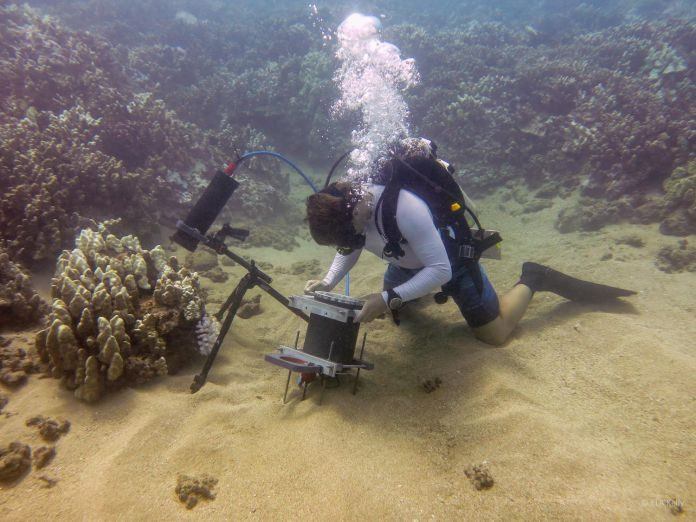
Researchers from Scripps Institution of Oceanography at the University of California San Diego have recently developed a new small imaging system. They have developed a diver-operated underwater microscope for analyzing millimetre-scale processes as they naturally occur on the seafloor. This microscope unveils the unseen view of the underwater world. This microscope is the first ever instrument that captures seafloor images at small scale.
Most organic processes in ocean arrive at small scales. When scientists remove organisms from their native habitats to study them in the lab, much of the information and its context are lost. To overcome this challenge, Jules Jaffe, Scripps oceanographer and his team developed a new type of underwater microscope. This microscope captures microorganisms with their natural activities without disturbing them.
Scientists develop this microscope with the aim of better understanding many ecological processes in underwater on a small scale. Researchers observed honeycomb pattern of initial algal colonization by using this microscope, which was previously unreported. They also identified growth in areas between the different coral polyps during coral bleaching.
The Benthic Underwater Microscope, or BUM, is a two-part system. This is like an underwater computer with a diver interface tied to a small imaging unit to analyse marine subjects at nearly micron resolution. This device consists of high magnification lens, a ring, glowing imaging abilities and flexibly tunable lens. The ring is fixed with LED lights for instant disclosure and lens is similar to the human eye for changing the view of the 3D structure.
Jaffe, head of the Jaffe Laboratory for Underwater Imaging at Scripps, “To understand the evolution of the dynamic processes taking place in the ocean, we need to observe them at the appropriate scale.”
Researchers used the imaging system while testing new technology’s ability to capture small-scale processes in underwater. This imaging system displays millimetre-sized coral polyps off the seashore of Israel in the Red Sea, and off Maui, Hawaii.
When experimenting in the red sea, scientists put up BUM for capturing interactions between two corals. These two corals are placed close to each other. The result came out with images, which unveils discharging of string. This is similar to filaments that hide enzymes from their stomach cavity to continue chemical turf battle for destroying tissue of other species in a competition for seafloor space.
Still, when the researchers placed corals of the same species next to each other, they did not eject these gastric fluids.
According to Mullen, “They can recognize friend versus foe.”
Additionally, scientists set up the instrument off Maui by following a largest coral bleaching events on record. It happens when a single cell of algae is present inside the coral polyp and eject themselves during high ocean temperature events.
According to scientists, “This microscope provides vision to the process of succession of algae. In this process, small filamentous algae is settled originally between coral polyps ridges and ultimately smother the living tissue. The images showed that algae is capable of strongly overflow living corals while bleaching process.”
Tali Treibitz, a former Scripps postdoctoral researcher, said, “This instrument is a part of a new trend in ocean research to bring the lab to the ocean, instead of bringing the ocean to the lab.”
Jaffe and Mullen are now preparing the instrument to take pictures of microscopic particles in the water near the coral’s surface to study how the flow of water over corals allows them to exchange the necessary gases to breathe.
