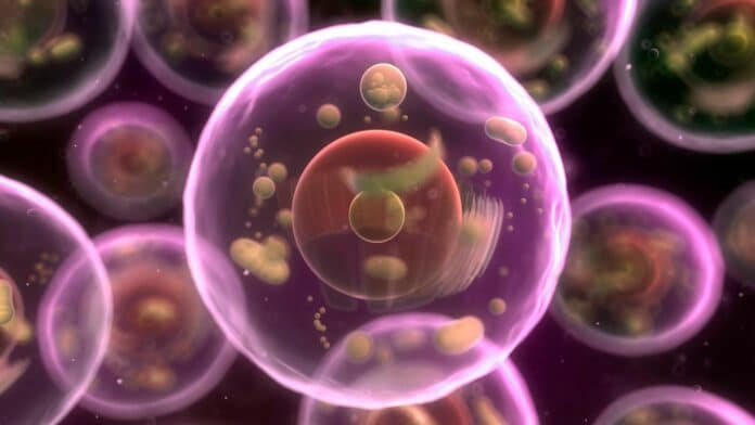
While nanoscale structural imaging of cells is now possible, a direct recording of the chemical composition of these domains is lacking. A novel technique was created by scientists at the Beckman Institute for Advanced Science and Technology to “see” the intricate details and chemical composition of a human cell with unparalleled clarity and precision. Their method approaches signal identification in a unique and counterintuitive way.
Rohit Bhargava, a professor of bioengineering at the University of Illinois Urbana-Champaign who led the study, said, “Now, we can see inside cells in a much finer resolution and with significant chemical detail more easily than ever. This work opens many possibilities, including a new way to examine the combined chemical and physical aspects that govern human development and disease.”
This new work is inspired from the last strides in chemical imaging.
Exposing a cell to IR light raises its temperature and leads to cell expansion. We can compare a poodle to a park bench to see that no two items absorb infrared wavelengths the same way. Night vision goggles also show that warmer objects generate stronger IR signatures than cooler ones. The same is true inside a cell, where several types of molecules release a particular chemical signature and absorb IR light at a different wavelength. Scientists can identify each one’s location by spectroscopically analyzing the absorption patterns.
Instead of analyzing the absorption patterns as a color spectrum, scientists interpreted the IR waves with a signal detector: a minute beam fastened to the microscope on one end, with a fine tip that scrapes the cell’s surface like the nanoscale needle of a record player.
After cell expansion, the motion of the signal detector becomes more exaggerated and generates “noise”: so-called static that impedes accurate chemical measurements.
Bhargava said, “It’s an intuitive approach because we are conditioned to think of larger signals as better. We think the stronger the IR signal, the higher a cell’s temperature becomes, the more it expands, and the easier it will be to see.”
Seth Kenkel, a postdoctoral researcher in Professor Bhargava’s lab and the study’s lead author, said, “It’s like turning up the dial on a staticky radio station — the music gets louder, but so does the static.”
“In other words, no matter how powerful the IR signal became, the quality of the chemical imaging could not advance.”
“We needed a solution to stop the noise from increasing alongside the signal.”
Instead of focusing their energies on the strongest possible IR signal, scientists began experimenting with the smallest signal they could manage, ensuring that they could effectively implement their solution before upping the strength.
Kenkel said, “Though “counterintuitive,” starting small allowed us to honor a decade of spectroscopy research and lay critical groundwork for the future of the field.”
The approach allows high-resolution chemical and structural imaging of cells at the nanoscale — a scale 100,000 times smaller than a strand of hair. Most importantly, this technique is free of fluorescent labeling or dyeing molecules to increase their visibility under a microscope.
Journal Reference:
- Seth Kenkel, Mark Gryka, et al. Chemical imaging of cellular ultrastructure by null-deflection infrared spectroscopic measurements. PNAS. DOI: 10.1073/pnas.2210516119
