
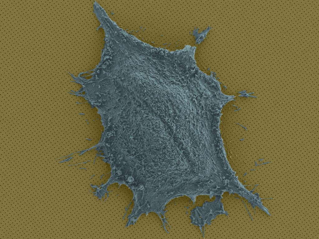
Immune response, metabolism, and cell-to-cell communication depend heavily on cell secretions such as proteins, antibodies, and neurotransmitters. The ability to predict the number of secretions without providing any information on when or where they are produced is a critical component of understanding cell secretions, which is essential for creating disease treatments.
Researchers at the University of Geneva and the BIOnanophotonic Systems Laboratory (BIOS) have created a revolutionary optical imaging technique that provides a four-dimensional view of cell secretions in space and time. They can map secretions as they are created while observing cell form and mobility by inserting individual cells into microscopic wells in a nanostructured gold-plated chip and then producing a phenomenon known as plasmonic resonance on the chip’s surface.
The scientists say their approach has “tremendous” promise for pharmaceutical development and basic research since it offers an unprecedentedly precise view of how cells function and interact. The technique allows scientists to screen cells individually in a high-throughput fashion.
The key component of the researchers’ technology is a 1 cm2 nanoplasmonic chip with hundreds of compartments for individual cells and millions of tiny holes. The chip is constructed of a gold substrate that has been nanostructured and is covered with a fine polymer mesh. A cell medium is placed inside each compartment to maintain the cells’ viability and health throughout imaging.
BIOS Ph.D. student and first author Saeid Ansaryan said, “Cell secretions are like the words of the cell: they spread out dynamically in time and space to connect with other cells. Our technology captures key heterogeneity regarding where and how far these ‘words travel.'”
A light beam causes the gold electrons to vibrate, which is how the nanoplasmonics part comes into play. Only particular wavelengths can pass through the nanostructure because of the way it was designed. The spectrum shifts when something on the chip’s surface, like protein secretion, changes the light that passes through. This shift is converted into variations in pixel intensity using a CMOS (Complementary Metal Oxide Semiconductor) image sensor and an LED.
Ansaryan said, “The beauty of our apparatus is that the nanoholes distributed across the entire surface transform every spot into a sensing element. This allows us to observe the spatial patterns of released proteins irrespective of cell position.”
The method has allowed scientists to glimpse two essential cellular processes – cell division and cell death – and to study delicate antibody-secreting human donor B-cells.
BIOS head Hatice Altug says, “We saw the cell content released during two forms of cell death, apoptosis, and necroptosis. In the latter, the content is released in an asymmetric burst, resulting in an image signature or fingerprint. This has never before been shown at the single-cell level.”
The cells under study can be quickly recovered since the procedure bathes the cells in a nourishing cell medium and does not require the poisonous fluorescent labels utilized by other imaging systems. As a result, the technique has great promise for use in developing pharmaceutical medications, vaccines, and other treatments. For instance, it might be used to assist researchers in understanding how cells react to various therapies individually.
Ansaryan said, “As the amount and pattern of secretions produced by a cell are a proxy for determining their overall effectiveness, we could also imagine immunotherapy applications where you screen patient immune cells to identify those that are most effective, and then create a colony of those cells.”
Journal Reference:
- Ansaryan, S., Liu, YC., Li, X. et al. High-throughput spatiotemporal monitoring of single-cell secretions via plasmonic microwell arrays. Nat. Biomed. Eng (2023). DOI: 10.1038/s41551-023-01017-1