Multiphoton microscope/two-photon excitation microscopy is a radiance imaging method. It permits imaging of living tissue up to about one millimeter in depth. Multiphoton Microscopy excitation, usually uses near infrared light. It also can accelerate fluorescent dyes. For each excitation, two photons of infrared light are absorbed. For minimizing the scattering in the tissue, it uses infrared light. Due to the absorption of multiphoton, the background signal is strongly suppressed. It increases the penetration depth for these microscopes.
Scientists have recently developed new multiphoton microscope and endoscope. This latest prototype has been designed by Imperil College, London in collaboration with the University of Bath. This new optical device could decrease the necessity of taking tissue samples during medical examinations and operations. Later, they are sending for testing to increase diagnosis and treatment. It could less down the healthcare costs.
The First optical device is a lightweight handheld microscope. It is designed to detect external tissue exposed while surgery. It can help physicians to compare normal and cancerous cells during operation. The advantage of this device that it can obtain high-quality 3D images of parts of the body while patients are doing the movements. For example, normal breathing. It is done by enabling to be applied to almost any exposed area of a patient’s body.
The second optical device is a tiny endoscope. It combines specially designed optical fibers and ultra-precise control of the light coupled into it. It can insert inside the body to bring out the internal cell scale examination, for example, during neurosurgery. It has the potential that it can provide high-resolution images by allowing surgeons to see inside individual cells at an adjustable depth below the surface of the tissue.
Presently, treatment of many diseases requires taking a sample of a tissue from the patient. After that, it tested in a laboratory by studying it under a microscope and then give back the results to the clinician.
Both new devices harness multiphoton excited fluorescence microscope technique to gather individual cells in their native tissues. It also could be useful for a consulting room or an operating theater to help physicians to detect diseased tissue and prescribe a rapid diagnosis.
Dr. Chris Dunsby of Imperial college London project leader, said, “These new devices open up existing possibilities in the field of intensity diagnose and could help to improve patient care in the future.”
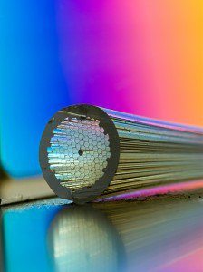
Both devices use new multicore optical fibers. This new multicore optical fiber was developed by Bath’s Centre for Photonics and Photonic material.
This handheld, lightweight microscope combines a tracking mechanism that makes restitution for the patient’s movement by ensuring the creation of fixed images. And the device called endoscope is a fraction of a millimeter in diameter. It doesn’t have any moving parts.
Professor Jonathan Knight, who led the University of Bath team, said, “This has been a very exciting project which has enabled us to develop fibers with a performance which we would have previously thought impossible.”
This new technique will be used by physicians for clinical use within next 5- 10 years.
